
Method and an apparatus for recording bladder volume

a technology of a bladder and a recording device, applied in the field of non-invasive methods and amplification devices for monitoring the bladder volume, can solve the problems of inability to accurately measure the volume of the bladder, the apparatus will inevitably generate mechanical noise, and the system is relatively large and heavy, so as to increase the transmission and reception efficiency of ultrasound signals

Inactive Publication Date: 2013-03-19
DANISH MEDICAL RES IVS
View PDF13 Cites 1 Cited by
  • Summary
  • Abstract
  • Description
  • Claims
  • Application Information

AI Technical Summary

Benefits of technology

[0018]The present invention now enables continuous monitoring of the volume of the bladder in humans or animals who or which move about free and easy or are at sleep. These continuous monitoring measurements of the bladder in humans or animals may be based on other areas of the bladder than the front and back wall. Also, these measurements are intended to increase the accuracy of the monitoring measurements of the bladder in humans or animals, as a means to minimize the likelihood of false measurements. The invention also provides an apparatus for monitoring the bladder volume which may be concealed from public view, on the body of the individual being monitored.

Problems solved by technology

The system, however, is relatively large and heavy because of the mechanical components used for the rotation of the ultrasound transducer and therefore not comfortable for the patients to wear.
Also, the bulky structure with the complicated, mechanical movements means that the apparatus will inevitably generate mechanical noise which may also be unpleasant to the patient.
The mechanically complicated structure will moreover be vulnerable to external impacts, which may occur if the unit is e.g. dropped.
The complicated structure will also be expensive to manufacture and require relatively great current maintenance costs.
Thus, it is a problem of the above mentioned techniques that they cannot be used in practice for long term monitoring of ambulatory patients, which is a great clinical need, e.g. in connection with the evaluation of the effect of recently developed medicine.
When the transmission source is placed above the center of the bladder, the front and back walls of the bladder can easily be detected using one single source, while the side walls, which may be close to parallel to the source, do not backscatter the sound waves optimally.
This system suffers from similar deficiencies as the above-mentioned apparatus, as the transducer elements are mounted in a single transducer, which means that the source of all scan planes originate in the transducer.
As the urinary bladder is an elastic non-symmetrical organ, which can be deformed by posture or by the quantity of urine contained in the bladder, the above mentioned measurement techniques may be prone to measure incorrectly as the measurements are based on front and back wall detection and the outlines of the bladder are estimated using these measurements.

Method used


Image

  • Method and an apparatus for recording bladder volume
  • Method and an apparatus for recording bladder volume
  • Method and an apparatus for recording bladder volume

Examples


Embodiment Construction

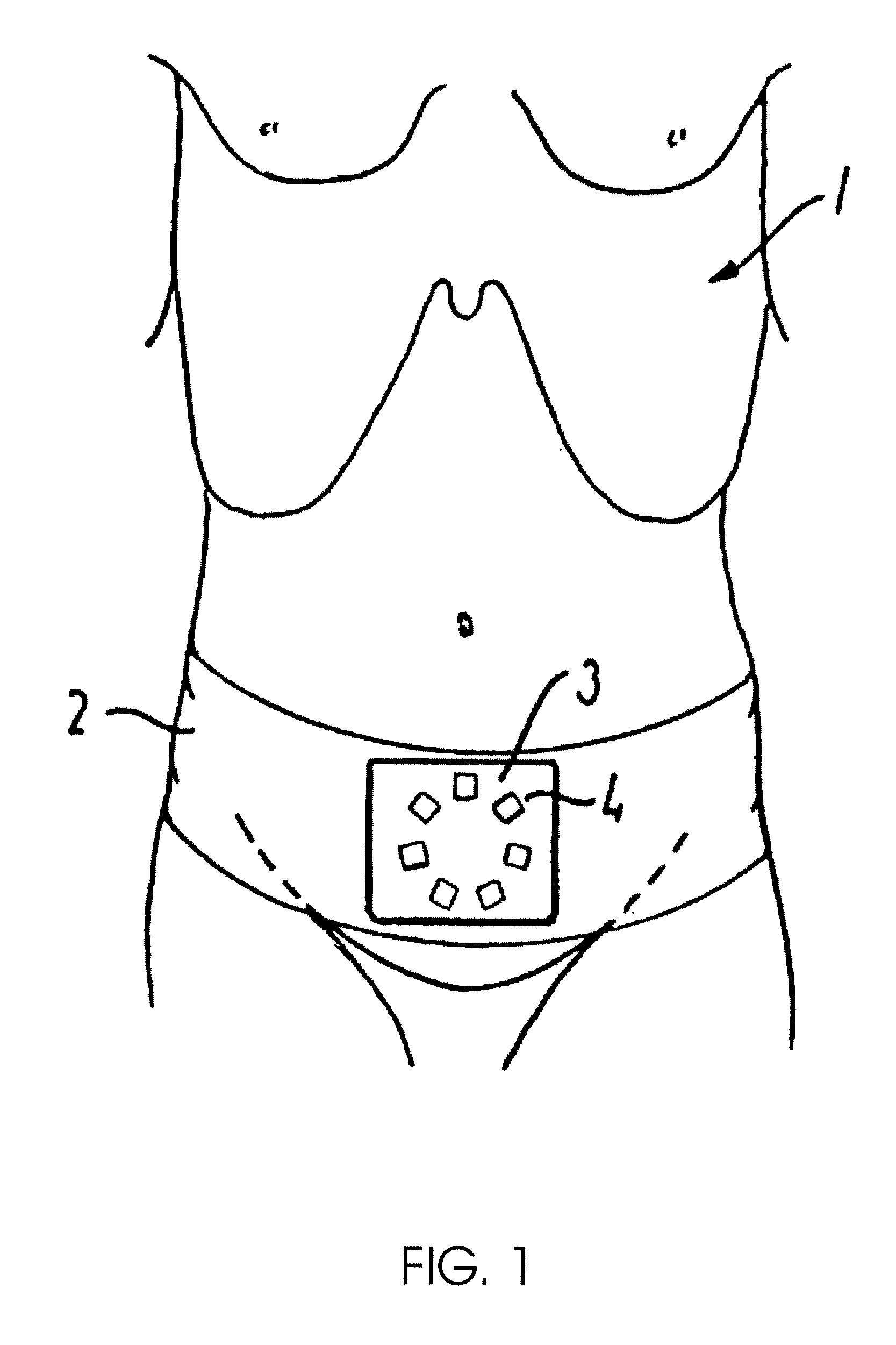
[0070]FIG. 1 shows the torso of an individual 1 who is to have his bladder volume monitored.

[0071]With this end in view, the individual is provided with a fixture in the form of a belt 2, which may also be integrated in the waistband of the pants. The apparatus 3 for the bladder monitoring measurement is arranged in the belt, based on a plurality of individual ultrasound transducer arrays 4 that are of the phased-array type.

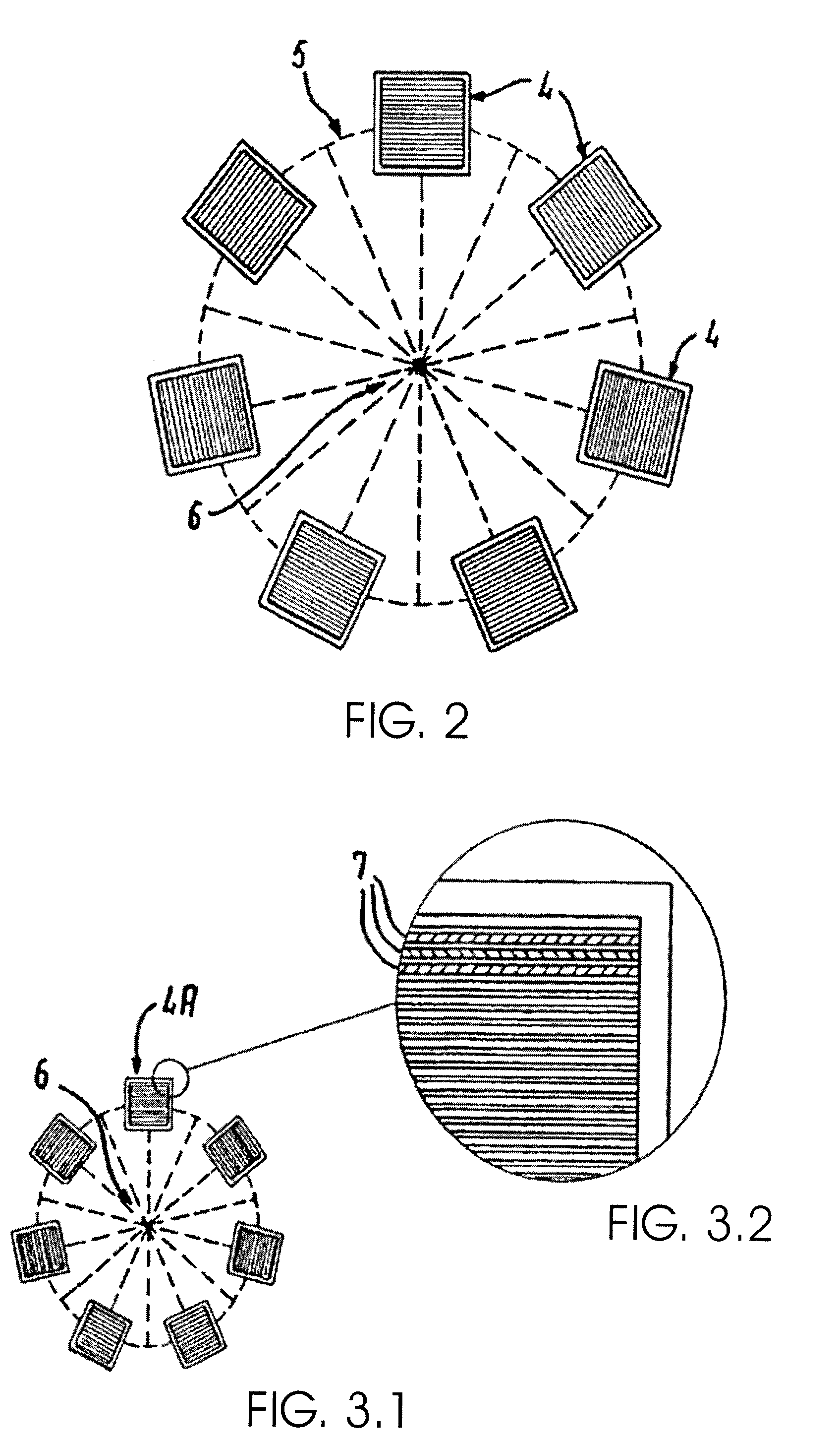
[0072]It is characteristic of the individual ultrasound transducer arrays that these can perform a scanning sweep in a plane without mechanical rotation of the individual transducer array, as the individual transducer array is composed of multiple piezoelectric crystals arranged in parallel which are capable of emitting signals in various angles by time delayed, individual excitation in a plane.

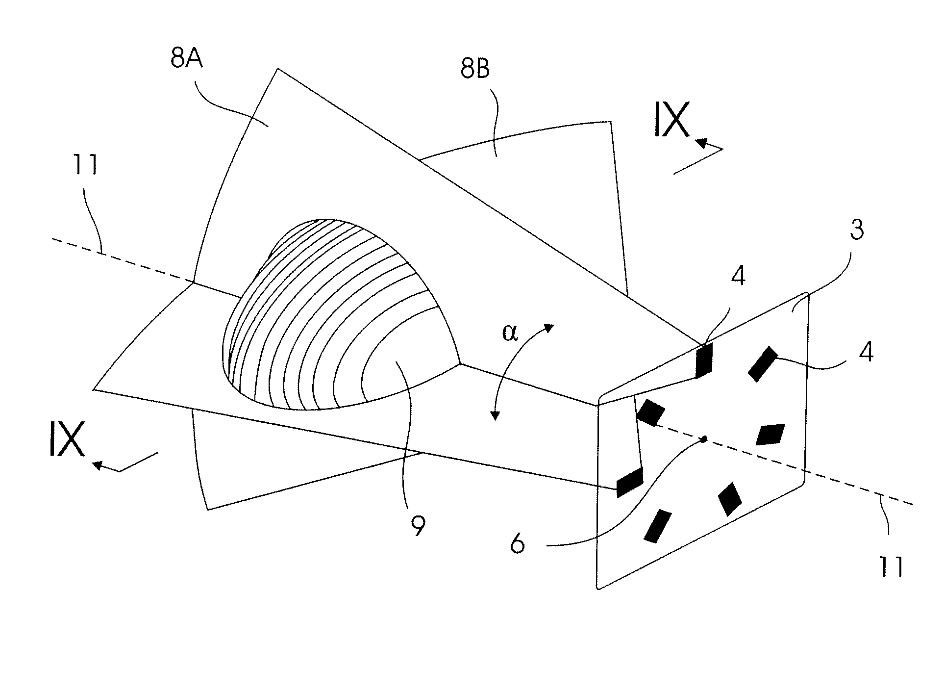
[0073]The fixture, in which the apparatus is mounted, is positioned such that an axis (as shown in FIG. 8) which extends from a center point of the apparatus through the m...


PUM


Abstract

A non-invasive method and apparatus for monitoring the bladder volume in humans or animals. The monitoring measurements are based on the analysis of ultrasound signals from at least two individual ultrasound transducer arrays. Each one individual ultrasound transducer array has a contact face, which is arranged in order to transmit an ultrasonic signal through an abdominal surface of an individual being monitored. The method and apparatus includes the arranging of each individual transducer array on the abdominal surface around an axis which extends from a point on the abdominal surface through a point on or within the urinary bladder. The method and apparatus further comprises that each individual transducer array is arranged to scan the bladder using a two-dimensional scan plane which extends radially from the transducer array through the bladder.

Description

CROSS-REFERENCE TO RELATED APPLICATIONS[0001]This application is a continuation-in-part of application Ser. No. 10 / 477,351 filed Jan. 9, 2004 now abandoned as the U.S. National State application under 35 U.S.C. 371 of International Application no. PCT / DK02 / 00320 filed May 16, 2002, the entire content of each of which is expressly incorporated herein by reference thereto.FIELD OF THE INVENTION[0002]The invention relates to a non-invasive method and apparatus for monitoring the bladder volume in humans or animals.BACKGROUND OF THE INVENTION[0003]The human or mammal urinary bladder is a triangularly shaped, hollow organ, located in the lower abdomen. The bladder walls are elastic, and can expand to hold the urine excreted from the kidneys and contract in order to empty the urine through the urethra. There are a number of disorders or ailments that can affect the urinary system, which could require the continuous monitoring of the bladder, long or short term, for diagnostic and / or treat...

Claims


Application Information

Patent Timeline
Patent Type & Authority Patents(United States)
IPC IPC(8): A61B8/00A61B5/20A61B8/08
CPCA61B5/202A61B5/204A61B8/0858A61B8/4488A61B8/4281A61B8/4227A61B8/4472A61B5/6808
Inventor KRISTIANSEN, NIELS KRISTIAN
Owner DANISH MEDICAL RES IVS
Learn More