
Crystalline composition of GM-CSF/GM-CSFR
- Summary
- Abstract
- Description
- Claims
- Application Information
AI Technical Summary
Benefits of technology
Problems solved by technology
Method used
Image

Examples
example 1
Atomic Coordinates of GM-CSF / GM-CSFR Complex
[0281]The atomic coordinates of the human GM-CSF / GM-CSFR crystalline complex are set forth in Table 1 (SEQ ID No. 15).
[0282]
Lengthy table referenced hereUS08489339-20130716-T00001Please refer to the end of the specification for access instructions.
example 2
Determination of Crystal Structure of GM-CSF / GM-CSFR Ternary Complex
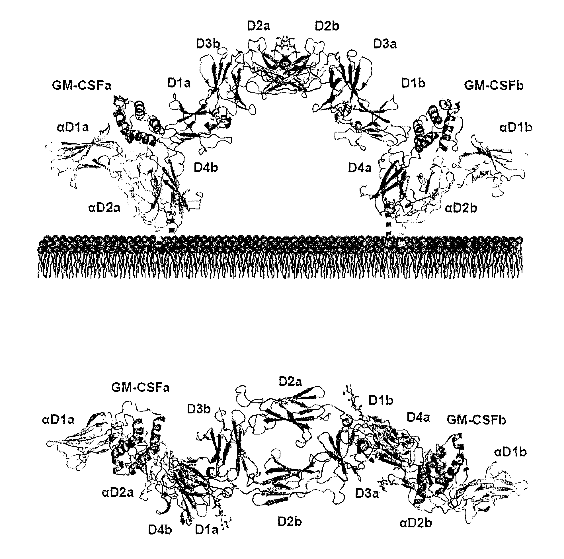
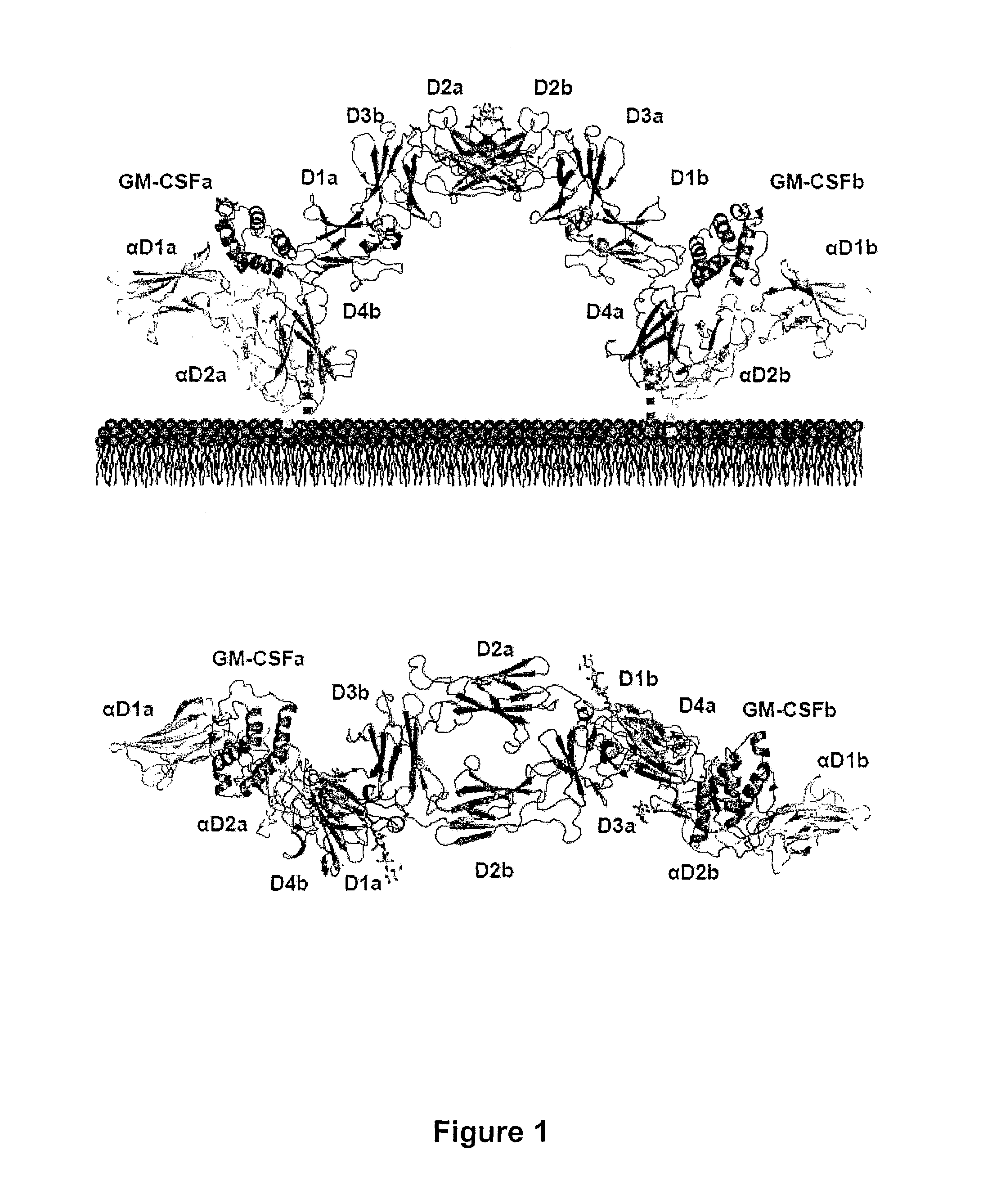
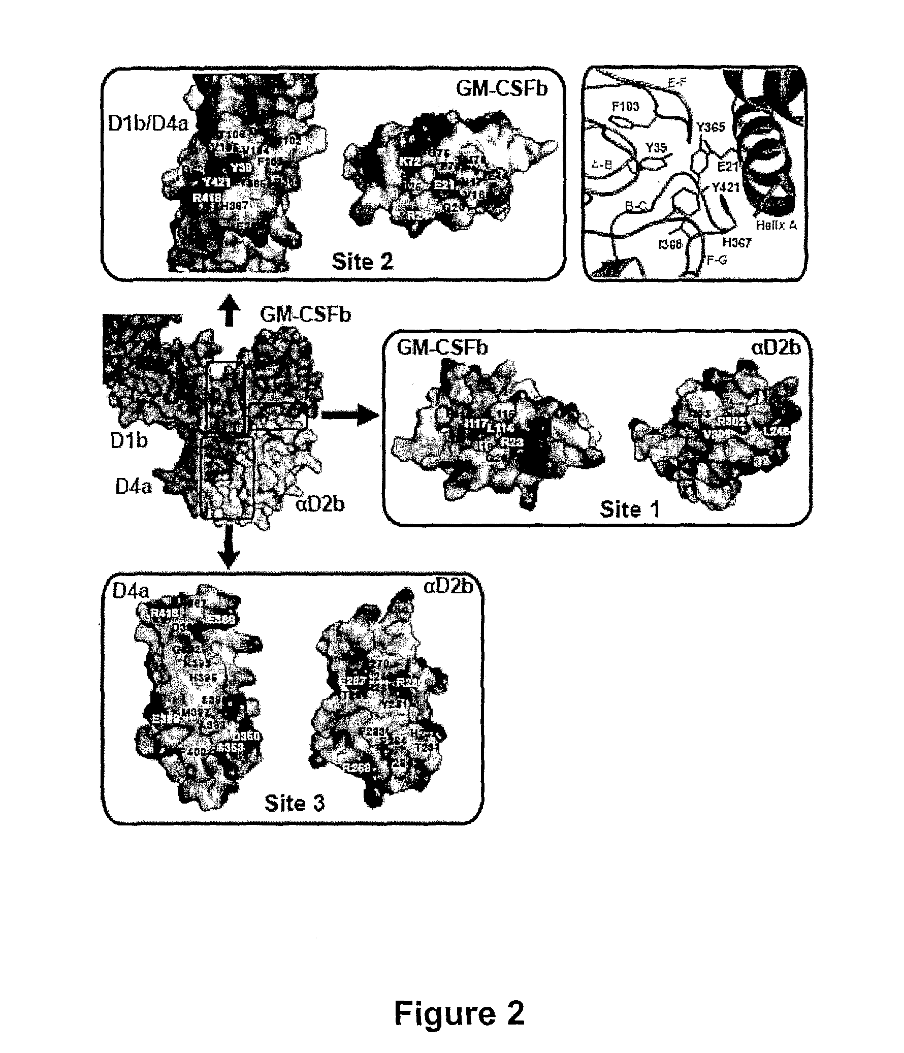
[0283]The crystal structure of the GM-CSF / GM-CSFR ternary complex is determined revealing a 2:2:2 hexamer consisting of two βc chains, two GM-CSFRα chains and two GM-CSF molecules (FIG. 1). The atomic coordinates are listed in Table 1 (Example 1). The structure adopted by GM-CSF in the ternary complex is similar to the structure of the isolated GM-CSF (Rozwarski et al, supra 1996). The same intertwined βc homodimer previously seen in the structure of the isolated βc subunit is also observed (Can et al, supra 2001). In the present case, the dimer is generated by a crystallographic twofold axis which runs through the center of the hexameric complex. Each βc chain consists of two cytokine receptor homology modules (CRMs), each of which consists of two fibronectin type III (FnIII) domains. A comparison of the βc chain in the complex compared to the published structure of the isolated molecule reveals that domain 4 has r...
example 3
Total Protein Concentration and Stoichiometry of the GM-CSF Ternary Complex
[0295]Formation and stoichiometry of the soluble ternary complex were characterized by a number of different techniques, usually at protein concentrations in the low micromolar range (McClure et al, supra 2003). For preparation of material used in crystallization studies, considerably higher protein concentrations (˜30 μM) are used. The fact that the stoichiometry of the crystallized ternary complex differs from what was observed in previous studies prompted an investigation into the impact of protein concentration on the stoichiometry of the GM-CSF ternary complex. Purified sβc (1.1-10.7 μM) was titrated alone or in the presence of equimolar sGM-CSFRα and a 1.5× molar excess of GM-CSF and fractionated the mixture by size exclusion chromatography (FIG. 6). The elution time of the ternary complex (TC) was markedly influenced by total protein concentration unlike the GM-CSFRα:GM-CSF binary complex (BC) or free ...
PUM
| Property | Measurement | Unit |
|---|---|---|
| Biological properties | aaaaa | aaaaa |
Abstract
Description
Claims
Application Information