Methods for treating melanoma
a technology for melanoma and treatment methods, applied in the field of antibodies and other agents, can solve the problems of hyperproliferation of tumor vasculature, drug effects that have little or no effect in patients with wild-type b-raf, and the difficulty of treating melanoma
- Summary
- Abstract
- Description
- Claims
- Application Information
AI Technical Summary
Benefits of technology
Problems solved by technology
Method used
Image
Examples
example 1
Evaluation of Melanomas for B-raf Mutations
[0205]A collection of xenografts have been established which are derived from patient melanoma tumors. The tumors were expanded by in vivo passage in NOD-SCID mice without any intervening in vitro cell culture. Genomic DNA samples were isolated from primary and passaged tumors using a Genomic DNA Extraction Kit (Bioneer Inc., Alameda, Calif.) following the manufacturers' instructions. The quality of the isolated DNA was checked by visualizing the DNA samples on a 1% agarose gel or a 0.8% E-Gel (Invitrogen Corporation, Carlsbad, Calif.). The DNA was confirmed to be intact by the presence of an approximately 20 kb size band with little or no visible degradation. The purified genomic DNA samples were sent to SeqWright Technologies, (Houston, Tex.) for nucleotide sequence analysis. The B-raf gene was obtained by amplifying genomic DNA samples with the Repli-G Mini Kit (Qiagen, Valencia, Calif.) followed by PCR amplification and purification. Th...
example 2
Inhibition of Melanoma Tumor Growth In Vivo by Anti-DLL4 Antibodies
[0208]NOD / SCID mice were purchased from Harlan Laboratories (Indianapolis, Ind.) and maintained under specific pathogen-free conditions and provided with sterile food and water ad libitum. The animals were housed in a U.S. Department of Agriculture-registered facility in accordance with NIH guidelines for the care and use of laboratory animals. The mice were allowed to acclimate for several days prior to the start of each study.
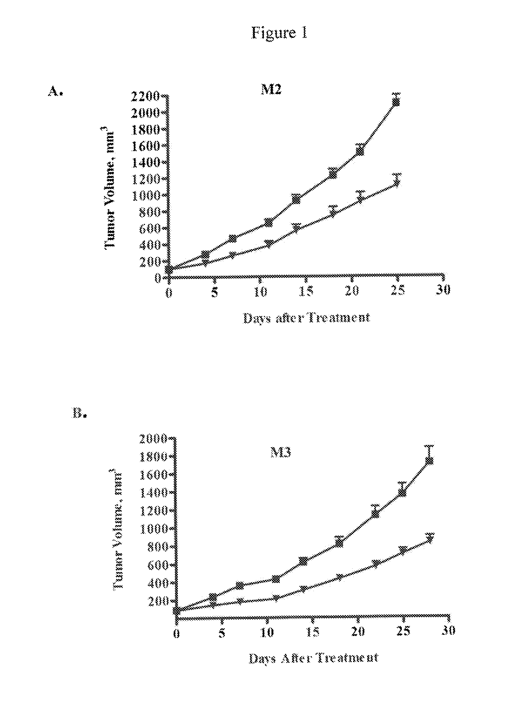
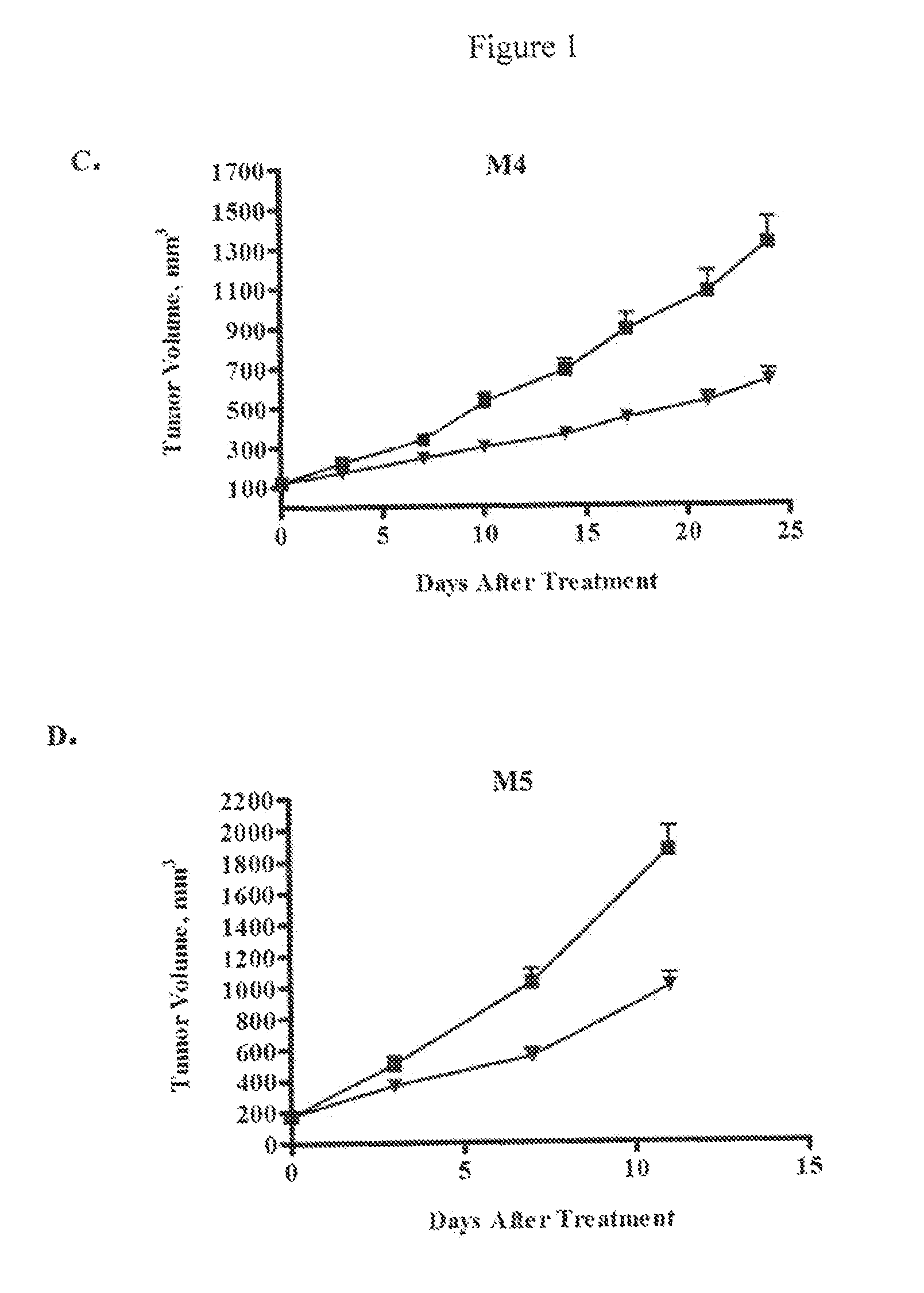
[0209]In general, tumor cells from a patient sample that have been passed as a xenograft in mice were prepared for injection into experimental animals. Tumor tissue was removed under sterile conditions, cut up into small pieces, minced completely using sterile blades, and single cell suspensions obtained by enzymatic digestion and mechanical disruption. Specifically, tumor pieces were mixed with ultra-pure collagenase III in culture medium and incubated at 37° C. for 1-4 hours. Digested cells ...
example 3
Inhibition of Melanoma Tumor Growth In Vivo by Anti-DLL4 Antibodies in Combination with a Chemotherapeutic Agent
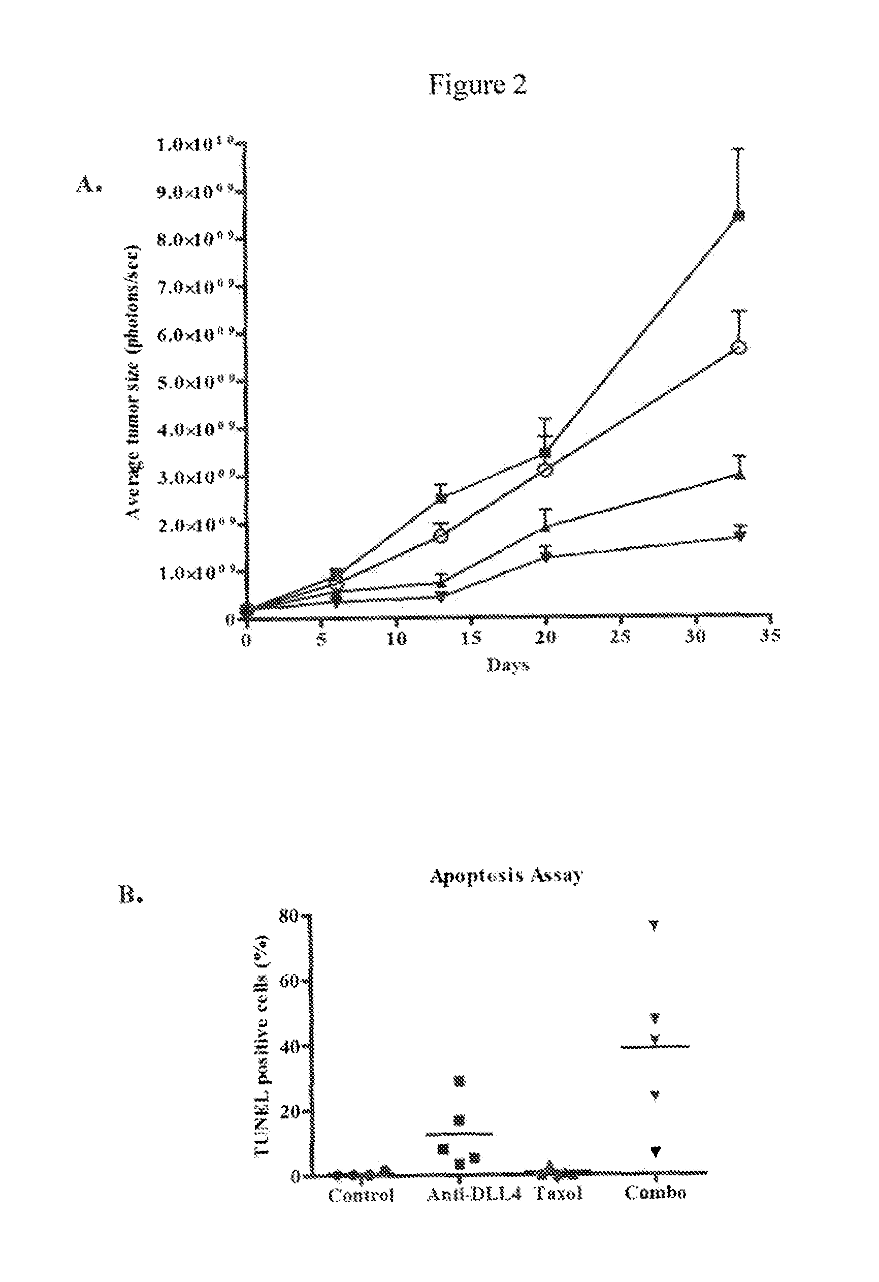
[0212]Luciferase-labeled M2 melanoma tumor cells (40,000 cells) were injected intradermally (orthotopic model) into 6-8 week old NOD / SCID mice. Tumor volumes were measured by determining the bioluminescent signal using an IVIS Imaging System (Caliper LifeSciences, Mountain View, Calif.). Tumors were allowed to grow until the average bioluminescent signal was approximately 2×10⁸ photons / sec. The animals were randomized into four groups (n=10 per group) and treated with a control antibody (anti-lysozyme antibody LZ-1, -▪-), an anti-DLL4 antibody (-▴-), taxol (-∘-), or a combination of taxol and anti-DLL4 antibody (-▾-). The anti-DLL4 antibody was a 1:1 mixture of anti-human DLL4 antibody and anti-mouse DLL4 antibody as described above. Antibodies were administered at 15 mg / kg once a week and taxol was administered at 10 mg / kg once a week. Both agents were administered intrap...
PUM
 Abstract
Description
Claims
Application Information
 - R&D
- Intellectual Property
- Life Sciences
- Materials
- Tech Scout
- Unparalleled Data Quality
- Higher Quality Content
- 60% Fewer Hallucinations
Browse by: Latest US Patents, China's latest patents, Technical Efficacy Thesaurus, Application Domain, Technology Topic, Popular Technical Reports.
© 2025 PatSnap. All rights reserved.Legal|Privacy policy|Modern Slavery Act Transparency Statement|Sitemap|About US| Contact US: help@patsnap.com


