Handheld dental camera and method for carrying out optical 3D measurement

a dental camera and optical 3d technology, applied in the field of handheld dental cameras for carrying out optical 3d measurement, can solve the problems of difficult to realize, limited depth measurement range and depth resolution, and insufficient economic utilization of the area sensor, so as to shorten the scanning procedure

Active Publication Date: 2013-11-05
SIRONA DENTAL SYSTEMS
View PDF25 Cites 112 Cited by
  • Summary
  • Abstract
  • Description
  • Claims
  • Application Information

AI Technical Summary

Benefits of technology

[0017]For this purpose, a polychromatic light source is used, of which the spectral range emitted in the form of the illuminating beam has a plurality of wavelengths. This illuminating beam is focused by means of a chromatic objective onto the object to be measured. Since a chromatic objective intensifies the effect of chromatic aberrations, the focal points for the different wavelengths of the illuminating beam are kept well apart. The focal points of the shortest and longest wavelengths of the spectral range of the illuminating beam can be spaced from each other by up to 5 mm, and they form the chromatic depth measurement range of the handheld dental camera. An elevation value can be assigned to each wavelength within this chromatic depth measurement range.
[0072]This scanning cycle omits the step in which the illuminating beam is moved back to its original position, so that the duration of the scanning procedure is shortened.

Problems solved by technology

Thus the depth measurement range and the depth resolution achievable in this application are limited.
Because of the shift of adjacent spectra, this technique has the reputation of being complicated for evaluation purposes, and the area of the area sensor is not utilized economically, because the filling factor is low.
However, the disadvantage of the chromatic confocal measuring method is that a spectral broadband light source must be used that has a wavelength spectrum that is as broad and continuous as possible.
These light sources are comparatively unwieldy and large, due to their design.
A compact light source such as a laser diode or a super-luminescent diode is less suitable for the chromatic confocal measuring method, since it typically has a rather narrow wavelength spectrum.
The depth measurement range is therefore greatly restricted and not suitable for measuring relatively large objects such as teeth.
Therefore, the classical scanning confocal measuring method suffers from the shortcoming that very large amounts of data accumulate that have to be processed for the purpose of carrying out good resolution.

Method used


Image

  • Handheld dental camera and method for carrying out optical 3D measurement
  • Handheld dental camera and method for carrying out optical 3D measurement
  • Handheld dental camera and method for carrying out optical 3D measurement

Examples


Embodiment Construction

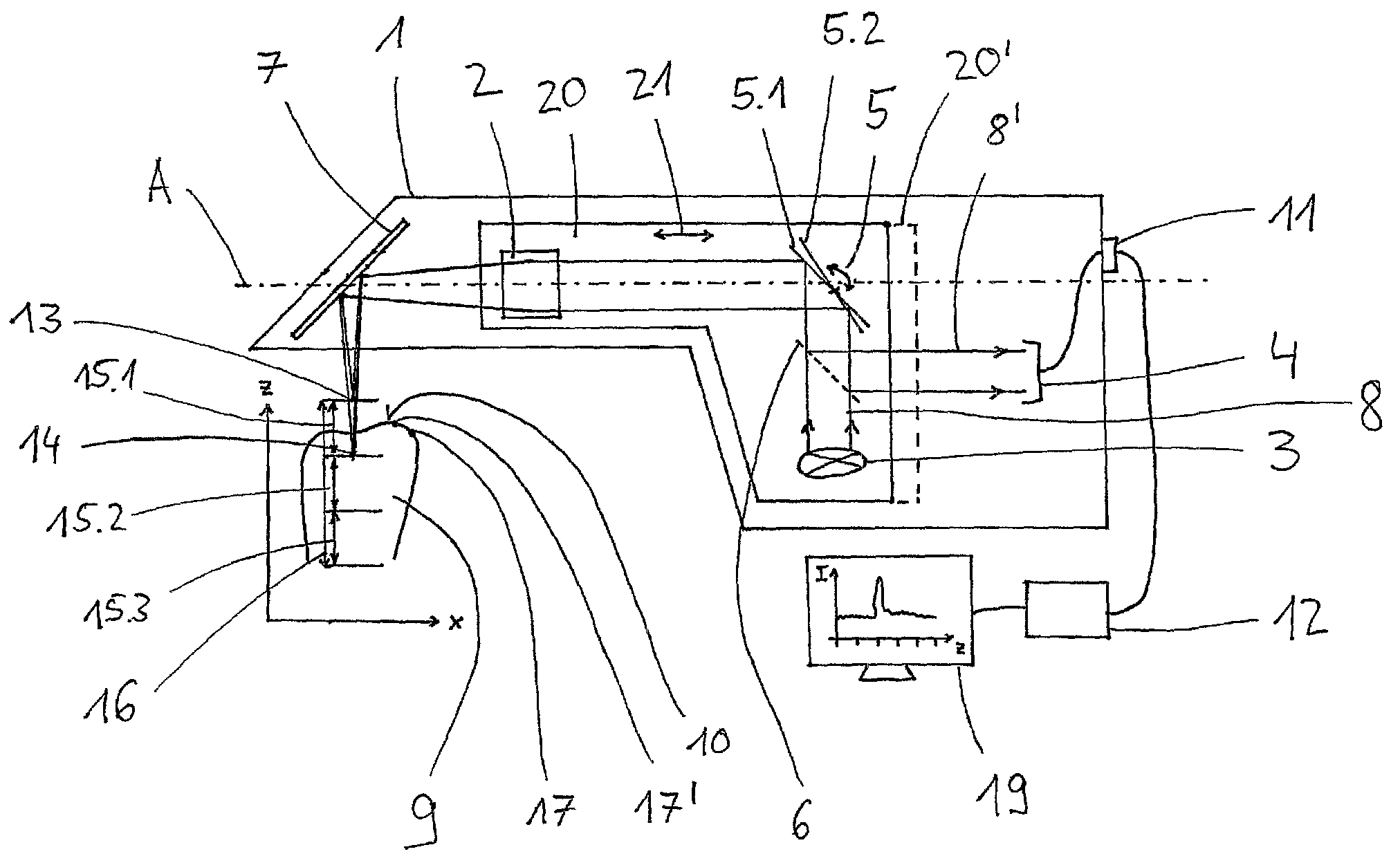
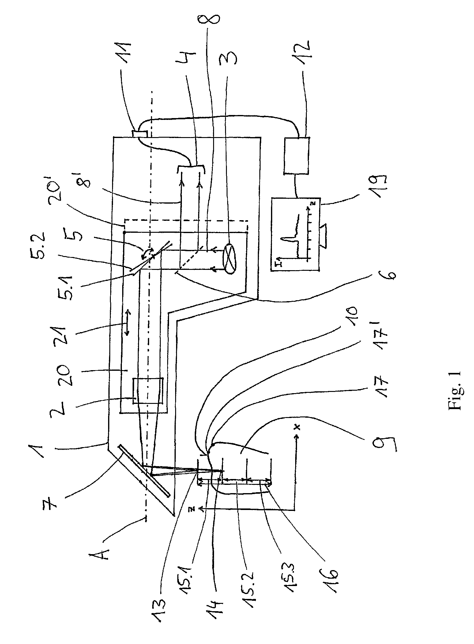
[0077]FIG. 1 shows an exemplary embodiment of a handheld dental camera 1 of the invention for carrying out 3D measurements. The handheld dental camera 1 comprises a scanning unit which, in this exemplary embodiment, comprises a chromatic objective 2, a polychromatic light source 3, a pivotal mirror 5, and a beam splitter 6, and which is capable of being moved along the longitudinal axis A within the handheld dental camera 1. The handheld dental camera 1 further comprises a color sensor 4, a deflector 7, for example a deflection mirror, and a connector 11 to which a data processing unit 12 can be connected.

[0078]The polychromatic light source 3 emits an illuminating beam 8 that passes through the beam splitter 6, for example, a semi-transparent mirror or a beam splitter prism, with as little obstruction as possible, and is deflected by the pivotal mirror 5 toward the chromatic objective 2. The illuminating beam 8 is focused by the chromatic objective and deflected by the deflector 7 ...


PUM


Abstract

A handheld dental camera performs three-dimensional, optical measurements. The camera includes a light source that emits an illuminating beam, a scanning unit, a color sensor, and a deflector. The scanning unit focuses the illuminating beam onto a surface of an object to be measured. The surface of the object reflects the illuminating beam and forms a monitoring beam, which is detected by the color sensor. Focal points of wavelengths of the illuminating beam form chromatic depth measurement ranges. The scanning unit stepwise displaces the chromatic depth measurement ranges by a step width smaller than or equal to a length of each chromatic depth measurement range, so that a first chromatic depth measurement range in a first end position of the scanning unit and a second chromatic depth measurement range in a second end position are precisely adjoined in a direction of a measurement depth, or are partially overlapped.

Description

RELATED APPLICATIONS[0001]This application is a continuation of International Application PCT / EP2010 / 052241, filed Feb. 23, 2010, claims benefit of the filing date of that application under 35 U.S.C. §120, and claims benefit under §119 of German Application No. 10 2009 001 086.6, filed Feb. 23, 2009. The entire contents of those two applications are incorporated herein by reference.TECHNICAL FIELD[0002]The invention relates to a handheld dental camera for carrying out optical 3D measurement using a confocal measuring method, which handheld dental camera comprises a chromatic objective, a polychromatic light source, and a color sensor, and also to a method for using the dental camera of the invention.PRIOR ART[0003]Confocal microscopy is well-known in the prior art and is disclosed, inter alia, in the patent specification U.S. Pat. No. 3,013,467.[0004]The chromatic confocal measuring method provides the possibility of effecting focusing without the need for mechanically moving compon...

Claims


Application Information

Patent Timeline
Patent Type & Authority Patents(United States)
IPC IPC(8): A61B1/04
CPCA61B5/0068A61B5/0088A61B5/1077G01B11/24G01B2210/50
Inventor THIEL, FRANK
Owner SIRONA DENTAL SYSTEMS
Why Eureka
Try Eureka