
Image processing apparatus and image processing method, and recording medium for processing breast image based on local contrast values in a local region in a mammary gland region of breast image

a mammary gland and image processing technology, applied in image analysis, image enhancement, instruments, etc., can solve the problems of insufficient viewability of (easiness to see) a local mammary gland structure and a lesion, insufficient accuracy of actual border between the mammary gland and the other tissue regions, and inability to accurately extract the mammary gland region. , to achieve the effect of stably calculated

Active Publication Date: 2013-12-03
FUJIFILM CORP
View PDF9 Cites 12 Cited by
  • Summary
  • Abstract
  • Description
  • Claims
  • Application Information

AI Technical Summary

Benefits of technology

The present invention provides an image processing apparatus and method that can improve the image quality of breast images by considering the contrast between mammary gland and fat regions, and the viewability of local mammary structure and lesions. The invention also ensures stable image processing results and prevents failures or variations in the extraction of mammary gland regions. This approach can enhance the accuracy and reliability of breast image processing and improve the diagnostic accuracy of breast cancer detection.

Problems solved by technology

However, with the above conventional arts, a global contrast is calculated on the basis of the signal values of the extracted mammary gland region as a whole, and there is a problem that viewability of (easiness to see) a local mammary gland structure and a lesion is not sufficiently considered.
However, a border between the mammary gland and the other tissue regions might be inaccurate in actuality, and there is a problem that the accurate extraction of the mammary gland region is extremely difficult.
Moreover, with the above conventional arts, since the contrast is calculated on the basis of the signal values only of the mammary gland region, the contrast with a peripheral region (fat region) is not sufficiently considered.

Method used


Image

  • Image processing apparatus and image processing method, and recording medium for processing breast image based on local contrast values in a local region in a mammary gland region of breast image
  • Image processing apparatus and image processing method, and recording medium for processing breast image based on local contrast values in a local region in a mammary gland region of breast image
  • Image processing apparatus and image processing method, and recording medium for processing breast image based on local contrast values in a local region in a mammary gland region of breast image

Examples


Embodiment Construction

[0055]The image processing apparatus and the image processing method according to the present invention will be described below in detail referring to the attached drawings.

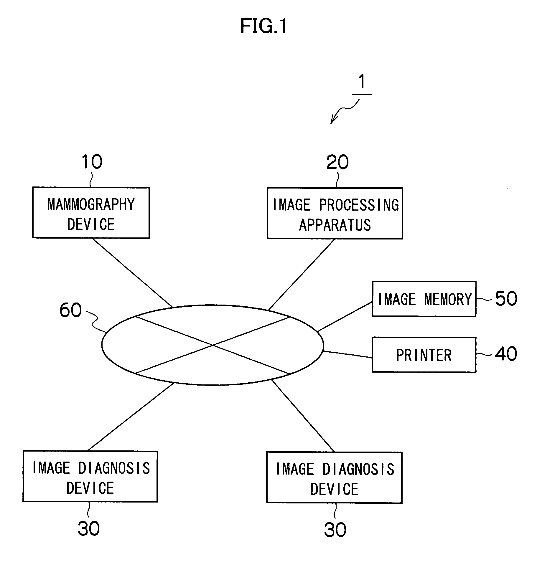
[0056]FIG. 1 is a diagram showing an entire configuration of an image diagnosis system to which an image processing apparatus of the present invention is applied.

[0057]As shown in FIG. 1, an image diagnosis system 1 comprises a mammography device 10 which picks up an image of a breast of a subject, an image processing apparatus 20 which applies image processing to a breast image (image data) photographed by the mammography device 10, an image diagnosis device 30 which displays an image to which image processing is applied by the image processing apparatus 20 for diagnosis, a printer (image output device) 40 which outputs the breast image photographed by the mammography device 10 or the image processed by the image processing apparatus 20 to a film and the like, and an image memory 50, which is a server in which i...


PUM


Abstract

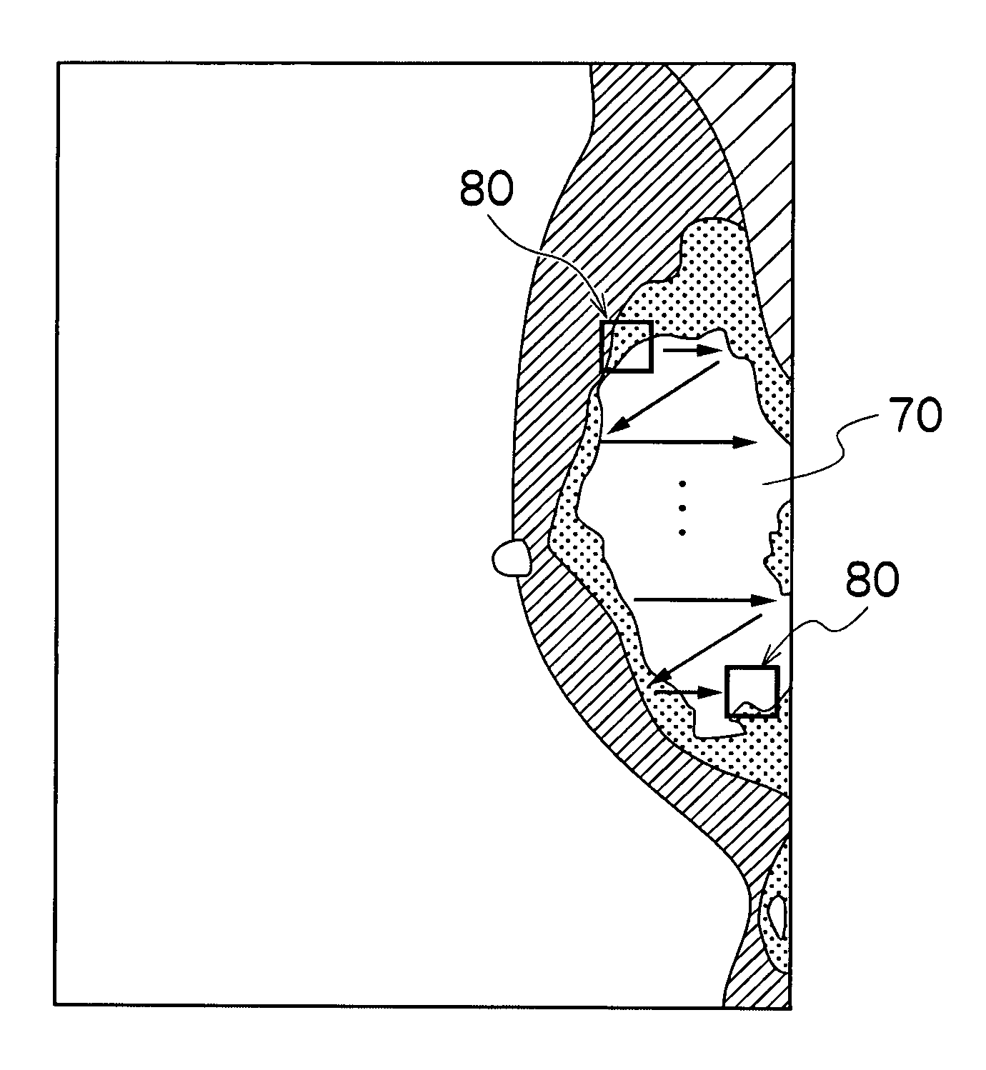
An image processing apparatus includes: an image obtaining device which obtains a breast image obtained by radiography of a breast; a mammary gland region extracting device which extracts a mammary gland region from the breast image; a local region setting device which sets a plurality of local regions around pixels belonging to the extracted mammary gland region; a local contrast value calculating device which calculates a plurality of local contrast values in a local regions, for each of the set plurality of local regions; and an image processing device which applies image processing to the breast image on the basis of the calculated plurality of local contrast values. Thus, considering a contrast between a mammary gland and a fat region, a stable image processing result can be obtained while enhancing viewability of a local mammary gland structure and a lesion.

Description

CROSS-REFERENCE TO RELATED APPLICATION[0001]This application claims priority under 35 USC 119 from Japanese Patent Application 2008-097888, filed Apr. 4, 2008, the disclosure of which is incorporated herein by reference.BACKGROUND OF THE INVENTION[0002]1. Field of the Invention[0003]The present invention relates to an image processing apparatus and an image processing method and particularly to a technology for obtaining a stable image processing result for a breast image with a large personal difference in mammography for breast diagnosis using an image of a breast photographed by X-ray.[0004]2. Description of the Related Art[0005]As a diagnosis using a medical image obtained by photographing a subject, mammography (breast X-ray photography) is conventionally known, for example. Breasts are different in size or mammary gland quantity among people and are said to be an organ with the largest personal difference in human tissues.[0006]Thus, features of the breast images taken by the ...

Claims


Application Information

Patent Timeline
Patent Type & Authority Patents(United States)
IPC IPC(8): G06K9/00
CPCG06T7/0081G06T5/008G06T2207/30068G06T2207/10116G06T7/11G06T5/94
Inventor MORITA, JUNYA
Owner FUJIFILM CORP
Learn More