
Retinal image analysis systems and methods

a retinal image and system analysis technology, applied in image enhancement, instruments, code conversion, etc., can solve the problems of difficult to quantify the above characteristics of retinal vessels on a large scale, high risk of severe visual loss requiring laser treatment, and labour-intensive presence of nvd in the ey

Inactive Publication Date: 2014-04-01
NAT UNIV OF SINGAPORE
View PDF16 Cites 18 Cited by
  • Summary
  • Abstract
  • Description
  • Claims
  • Application Information

AI Technical Summary

Benefits of technology

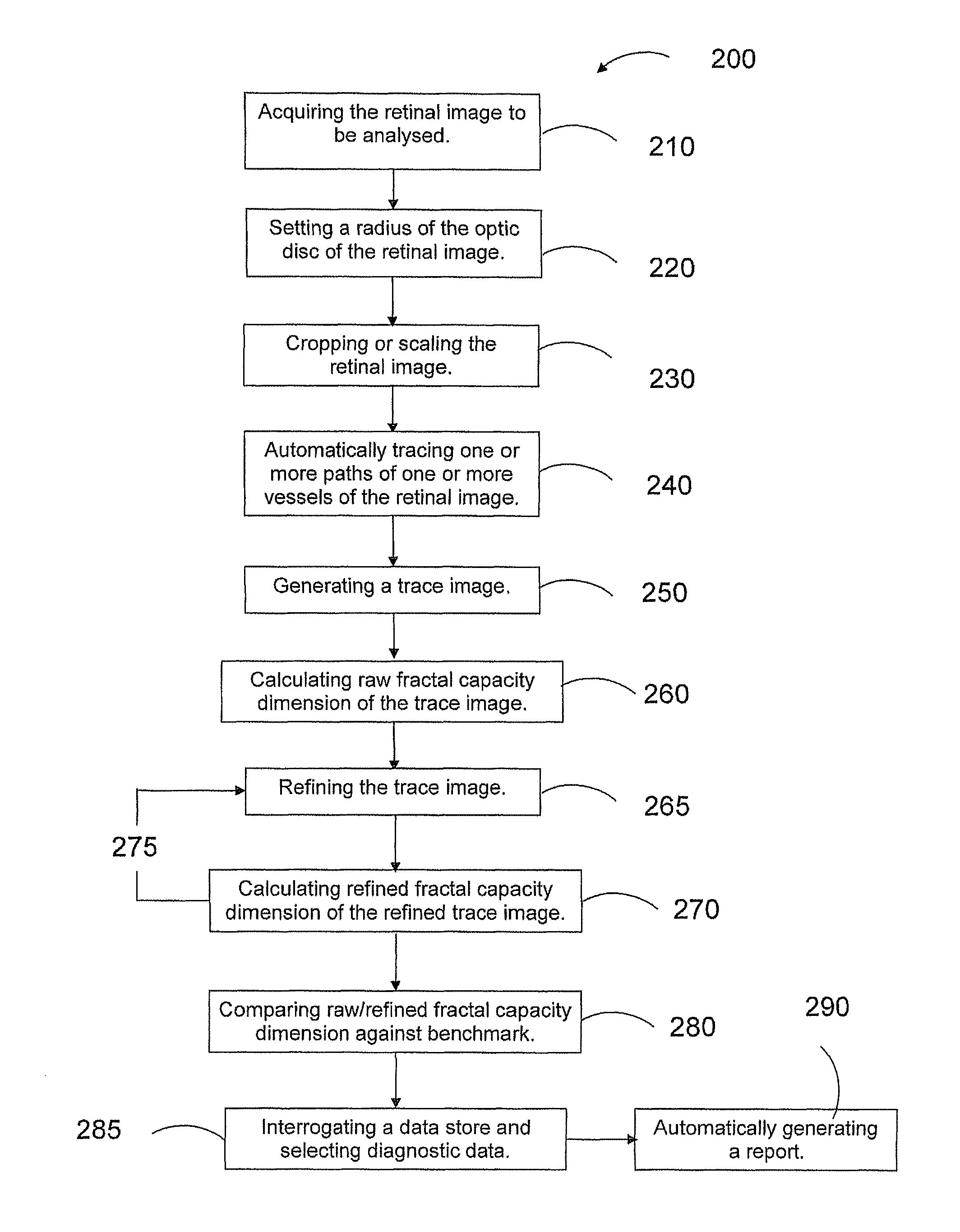
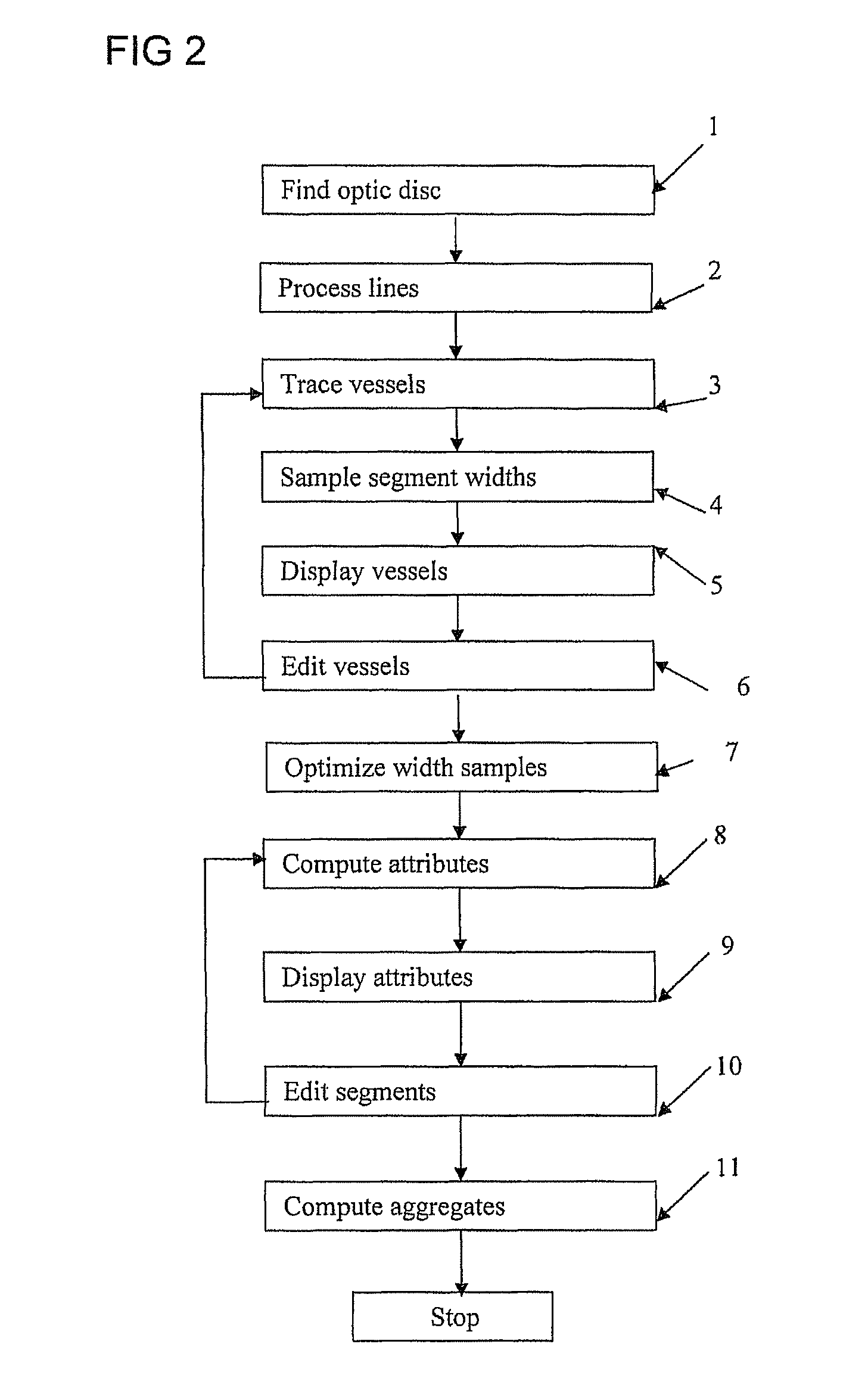
The patent proposes an automated system and method for analyzing retinal images to extract multiple characteristics. These characteristics can then be used to predict cardiovascular risk or diagnose specific cardiovascular conditions. The system is designed to extract information from large numbers of retinal images with high reliability. The method includes cropping or scaling the retinal image to improve image quality and facilitate comparisons.

Problems solved by technology

However, it is difficult to quantify the above characteristics of retinal vessels on a large scale as the process would involve repeated measurements of the diameters of the arterioles and venules in the retinal images by trained human graders.
This is labour intensive and the results can vary when different human graders are used.
The presence of NVD in an eye is a high risk characteristic for severe visual loss requiring laser treatment.

Method used


Image

  • Retinal image analysis systems and methods
  • Retinal image analysis systems and methods
  • Retinal image analysis systems and methods

Examples


Embodiment Construction

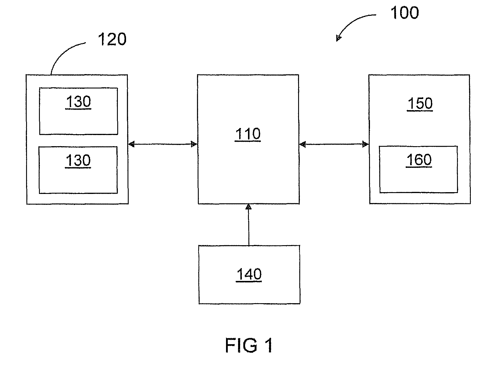
[0087]Referring to FIG. 1, there is provided a diagnostic retinal image system 100 in accordance with embodiments of the present invention. The system 100 comprises a processor 110 coupled to be in communication with an output device 120 in the form of a pair of displays 130 according to preferred embodiments of the present invention. The system 100 comprises one or more input devices 140, such as a mouse and / or a keyboard and / or a pointer, coupled to be in communication with the processor 110. In some embodiments, one or more of the displays 130 can be in the form of a touch sensitive screen, which can both display data and receive inputs from a user, for example, via the pointer.

[0088]The system 100 also comprises a data store 150 coupled to be in communication with the processor 110. The data store 150 can be any suitable known memory with sufficient capacity for storing configured computer readable program code components 160, some or all of which are required to execute the pre...


PUM


Abstract

A platform is proposed for automated analysis of retinal images, for obtaining from them information characterizing retinal blood vessels which may be useful in forming a diagnosis of a medical condition. A first aspect of the invention proposes that a plurality of characteristics of the retina are extracted, in order to provide data which is useful for enabling an evaluation of cardiovascular risk prediction, or even diagnosis of a cardiovascular condition. A second aspect uses fractal analysis of retinal images to provide vascular disease risk prediction, such as, but not limited to, diabetes and hypertension.

Description

RELATED APPLICATION[0001]This application claims priority to PCT application PCT / SG2009 / 000040 filed Feb. 3, 2009, which claims priority to U.S. Provisional Patent Application No. 61 / 123,360, filed Apr. 8, 2008.FIELD OF THE INVENTION[0002]The present invention relate to systems and methods for analyzing retinal images, and for obtaining from them information characterizing retinal blood vessels which may be useful in forming a diagnosis of a medical condition.BACKGROUND TO THE INVENTION[0003]Existing non-invasive methods for monitoring cardiovascular disorders utilize blood pressure (see for example U.S. Pat. Nos. 5,961,467, 4,669,485, 5,365,924, 6,135,966, 5,634,467, 5,178,151). However, clinical studies have provided indications that changes in retinal vasculature (e.g., narrowing of retinal arterioles and widening of retinal venules) may be an early indicator of cardiovascular disease (CVD) and other conditions, such as hypertension and diabetes. The conditions of retinal arterio...

Claims


Application Information

Patent Timeline
Patent Type & Authority Patents(United States)
IPC IPC(8): G06K9/00
CPCA61B3/12G06T2207/30101G06K2009/00932G06T2207/30041G06T2207/20072G06K9/0061G06T7/0012G06V40/193G06V40/14
Inventor HSU, WYNNELEE, MONG LIWONG, TIEN YIN
Owner NAT UNIV OF SINGAPORE
Learn More