Device and method for generating soft tissue contrast images

a technology of contrast image and soft tissue, which is applied in the field of generating soft tissue contrast image, can solve the problems of not all patients being allowed to be imaged, patient cannot be imaged by mri procedure, and soft tissue discrimination, so as to achieve fast image acquisition, increase the resolution of ct image, and broaden the effect of x-ray beams

Inactive Publication Date: 2014-04-29
KONINK PHILIPS ELECTRONICS NV
View PDF17 Cites 7 Cited by
  • Summary
  • Abstract
  • Description
  • Claims
  • Application Information

AI Technical Summary

Benefits of technology

[0022]For instance, in an embodiment information of the x-ray absorption distribution within the area of interest is obtained from detected x-ray signals, whereas according to another embodiment information of the location of the examination object is obtained from the detected x-ray signals. The latter information is preferably used in the model by setting the conductivity distribution, the x-ray absorption distribution and the efficiency distribution outside the examination object to zero which much reduces the calculation efforts for reconstructing soft tissue images.
[0023]According to another embodiment, an information of the location of the electromagnetic signal receiver, in particular of signal receiver elements, with respect to the area of interest is generated and used in the model. All these embodiments finally lead to a reduction of computation time and to an improvement of the signal quality of the soft tissue contrast images.
[0028]According to another preferred embodiment in which the x-ray source unit comprises a pulsed x-ray source and a collimator unit, the collimator unit comprises one or more controllable collimator elements, each of said collimator elements being provided for allowing a pencil beam to pass through in a first control state and for blocking an incident pencil beam in a second control state, and a collimator control unit for controlling said controllable collimator element. Generally, according to the present invention one or more pencil beams, i.e. a low number of pencil beams, are simultaneously incident on the examination object. This, however, has the disadvantage that a large amount of the x-ray radiation generated by a conventional x-ray source is blocked. However, to enable a reasonable signal to noise ratio, a certain threshold energy has to be introduced into the examination object per x-ray pulse. Hence, according to this preferred embodiment, not only a single or a low number of pencil beams is used but a larger number of pencil beams is made to be incident onto the examination object simultaneously.
[0037]The resolution of the CT image can be higher than the soft tissue image as the pencil beam x-ray photons may be recorded using several detectors. It is also generally possible to broaden the x-ray beams and use the device according to the present invention in a conventional CT mode with fast image acquisition.
[0038]The pencil beam like acquisition of the signals allows that CT acquisition virtually without blurring due to scattered radiation. Moreover, the scattered intensity may be recorded, adding information about the tissue, i.e. quantification of photo- and Compton-effect.
[0039]Hence, the main advantages of the present invention are that a higher resolution soft tissue contrast image can be generated which adds only negligible extra radiation to the examination. No contrast medium is needed. The device may also be used to deliver high quality classic CT images.

Problems solved by technology

One of the weak points of CT is, however, the discrimination of soft tissues.
Magnetic resonance imaging (MRI) has an excellent soft tissue contrast, but not all patients are allowed to be imaged by an MRI procedure.
For instance when the patient has some sort of implants, such as a pacemaker, a patient cannot be imaged by an MRI procedure.

Method used


Image

  • Device and method for generating soft tissue contrast images
  • Device and method for generating soft tissue contrast images
  • Device and method for generating soft tissue contrast images

Examples


first embodiment

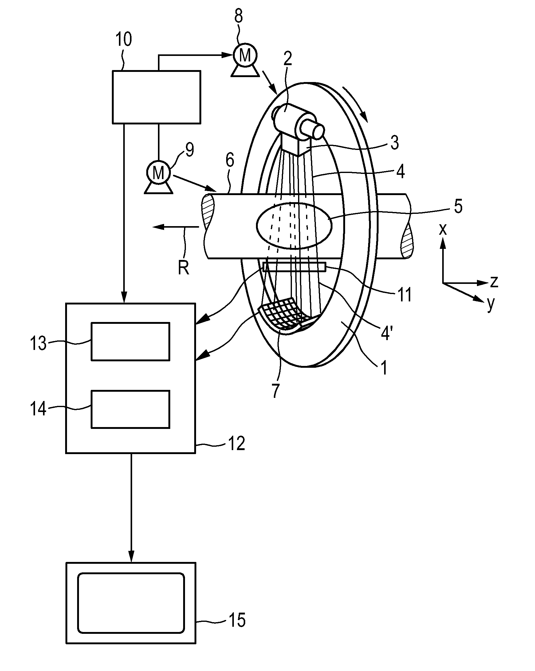
[0045]FIG. 1 shows a device according to the present invention. In this embodiment the mechanical layout is similar to a conventional CT imaging device. The device comprises a gantry 1 which is capable of rotating about an axis of rotation R which extends parallel to the z direction. A radiation source unit including an x-ray source 2, such as an x-ray tube, is mounted on the gantry 1. The x-ray source unit further comprises a collimator unit 3 which forms a bundle of pencil beams 4 (at least one pencil beam) from the radiation beam emitted by the x-ray source 2.

[0046]The pencil beams traverse a (symbolically shown) object 5, such as a patient, in an area of interest within a cylindrical examination zone 6. After having traversed the examination zone 6, the portion of the x-ray radiation 4′ of the pencil beams, that has not been absorbed by the object 5, is incident on an x-ray detector unit 7, in this embodiment a two-dimensional energy-resolving detector, which is also mounted on ...

third embodiment

[0089]FIG. 3 shows a device according to the present invention. According to this embodiment, the mechanical layout of the device is similar to the layout of an x-ray device applied for tomosynthesis. The device comprises a C-arm 20, the ends of which accommodate the x-ray source 2 and the x-ray detector 7. The C-arm 20 is suspended from an L-arm 21, by way of a pivot 22, so as to be rotatable about the horizontal propeller axis 23. The L-arm 21 is suspended from a displaceable carriage 24 by way of a further pivot 25. Said carriage 24 is suspended from the ceiling 26. The pivot 25 enables rotation about the vertical axis 27. The L-arm 21 can be displaced in the horizontal direction by way of the carriage 24. The object 5 to be examined which is symbolically shown is arranged on a table 28, which is mounted on a base 29 whose height can be adjusted and which is also displaceable in the horizontal direction 30. For acquisition of electromagnetic signals an electromagnetic signal rece...

fourth embodiment

[0099]FIG. 4 schematically shows a device according to the present invention. In addition to the embodiment shown in FIG. 2 this embodiment comprises acoustic signal receivers 31, in particular a plurality of microphones that are arranged close to the object 5. These acoustic signal receivers 31 are adapted for receiving acoustic signals from the object 5.

[0100]The tissue is heated when x-ray radiation is incident. Hence, the tissue expands and emits an acoustic pulse which can be measured by the acoustic signal receivers 31. The signal processor unit 12 is, according to this embodiment, adapted for also processing said acoustic signals and for reconstructing a separate soft tissue contrast image of the area of interest within the object 5 by use of an appropriate model, which preferably uses as model parameters the x-ray absorption distribution, thermal expansion, compressibility, density, acoustic velocity and / or acoustic absorption. Hence, some additional information to the tissu...


PUM


Abstract

The present invention relates to a device for generating soft tissue contrast images of an area of interest of an examination object (5) comprising soft tissue. The proposed device comprises: an x-ray source unit (2, 3) for emitting one or more pulsed pencil x-ray beams (4), an actuator (8, 9) for actuating said x-ray source unit (2, 3) along and / or around said area of interest to direct said one or more pencil beams (11) onto said area of interest from various directions, an electromagnetic signal receiver (11) for receiving electromagnetic signals from the area of interest resulting from absorption and scatter of x-ray photons within the area of interest, and a signal processor unit (12) for processing said received electromagnetic signals and reconstructing a soft tissue contrast image of the area of interest.

Description

FIELD OF THE INVENTION[0001]The present invention relates to a device and a corresponding method for generating soft tissue contrast images of an area of interest of an examination object comprising soft tissue.BACKGROUND OF THE INVENTION[0002]Computed tomography (CT) generates high-quality images of bone-structures and contrast agent distributions. One of the weak points of CT is, however, the discrimination of soft tissues. Magnetic resonance imaging (MRI) has an excellent soft tissue contrast, but not all patients are allowed to be imaged by an MRI procedure. For instance when the patient has some sort of implants, such as a pacemaker, a patient cannot be imaged by an MRI procedure.SUMMARY OF THE INVENTION[0003]It is an object of the present invention to provide a device and a corresponding method for generating soft tissue contrast images of an area of interest of an examination object, such as a patient, comprising soft tissue, which can also be applied to patients which cannot...

Claims


Application Information

Patent Timeline
Patent Type & Authority Patents(United States)
IPC IPC(8): G06K9/00
CPCA61B6/4241A61B6/482A61B6/4021A61B6/484A61B6/483
Inventor GLEICH, BERNHARD
Owner KONINK PHILIPS ELECTRONICS NV
Why Eureka
Try Eureka