Method and apparatus for dual-modality ultrasonic and nuclear emission mammography

a nuclear emission and ultrasonic technology, applied in the field of breast imaging, can solve the problems of reducing the sensitivity of mammography with increasing mammographic density, affecting the accuracy of mammography, and a less than perfect screening method of mammography

Active Publication Date: 2014-10-07
MAYO FOUND FOR MEDICAL EDUCATION & RES
View PDF12 Cites 19 Cited by
  • Summary
  • Abstract
  • Description
  • Claims
  • Application Information

AI Technical Summary

Benefits of technology

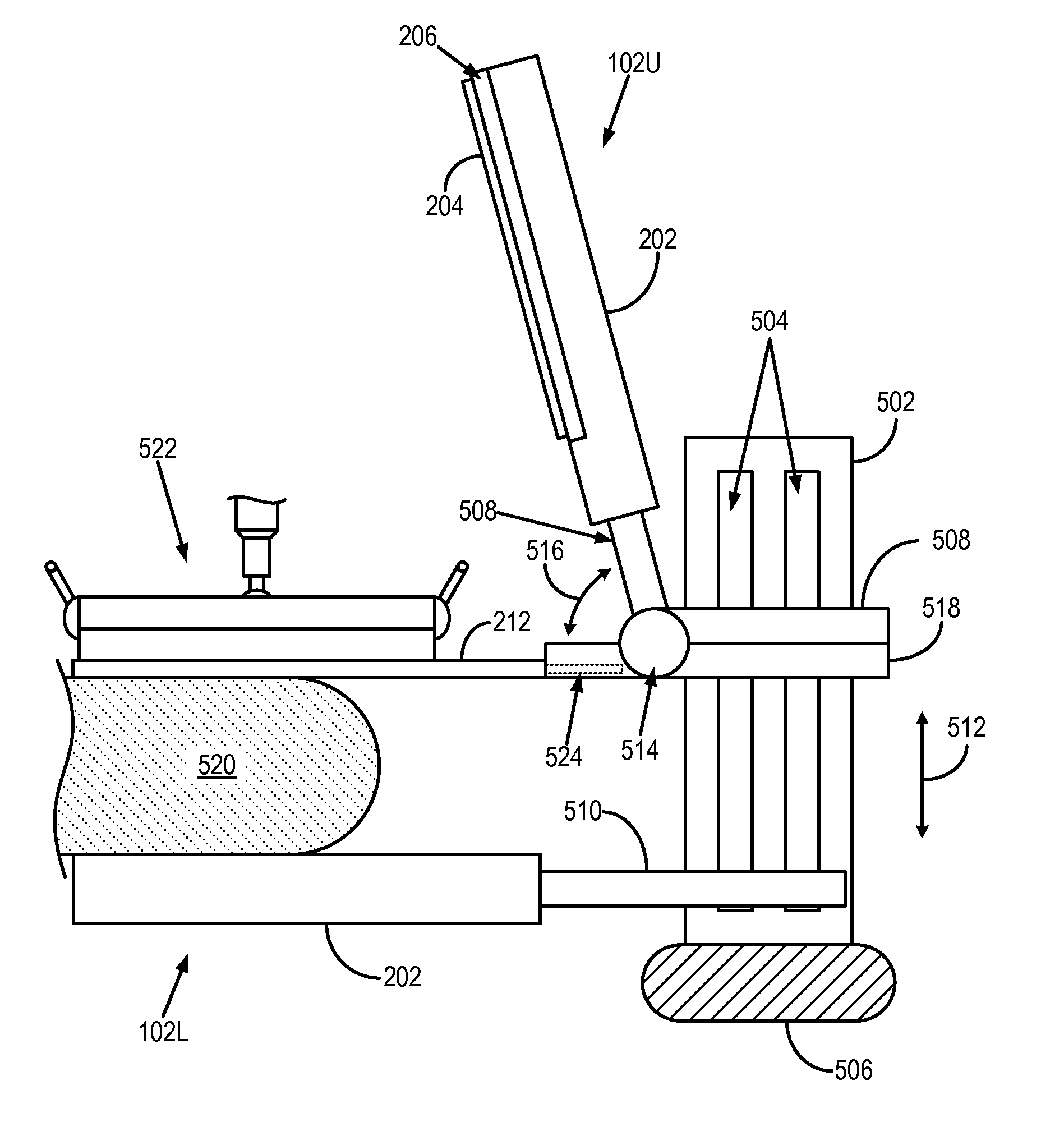
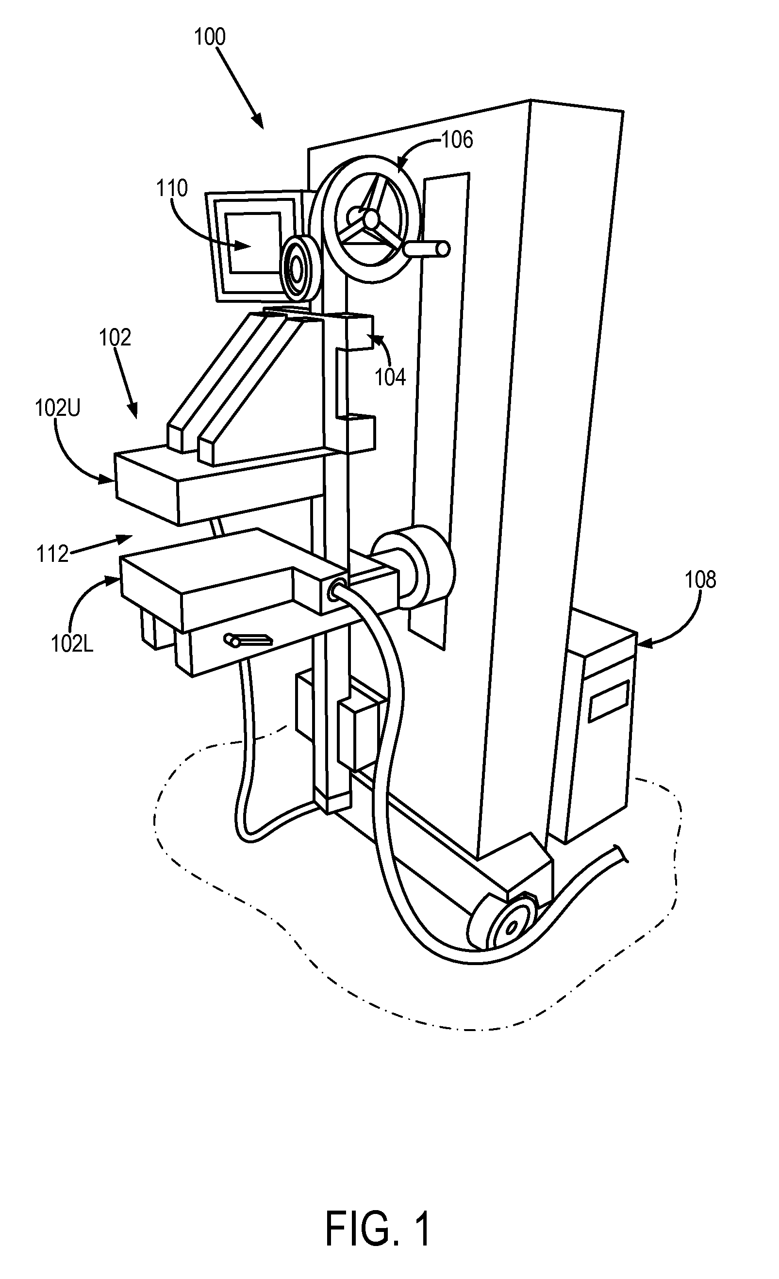
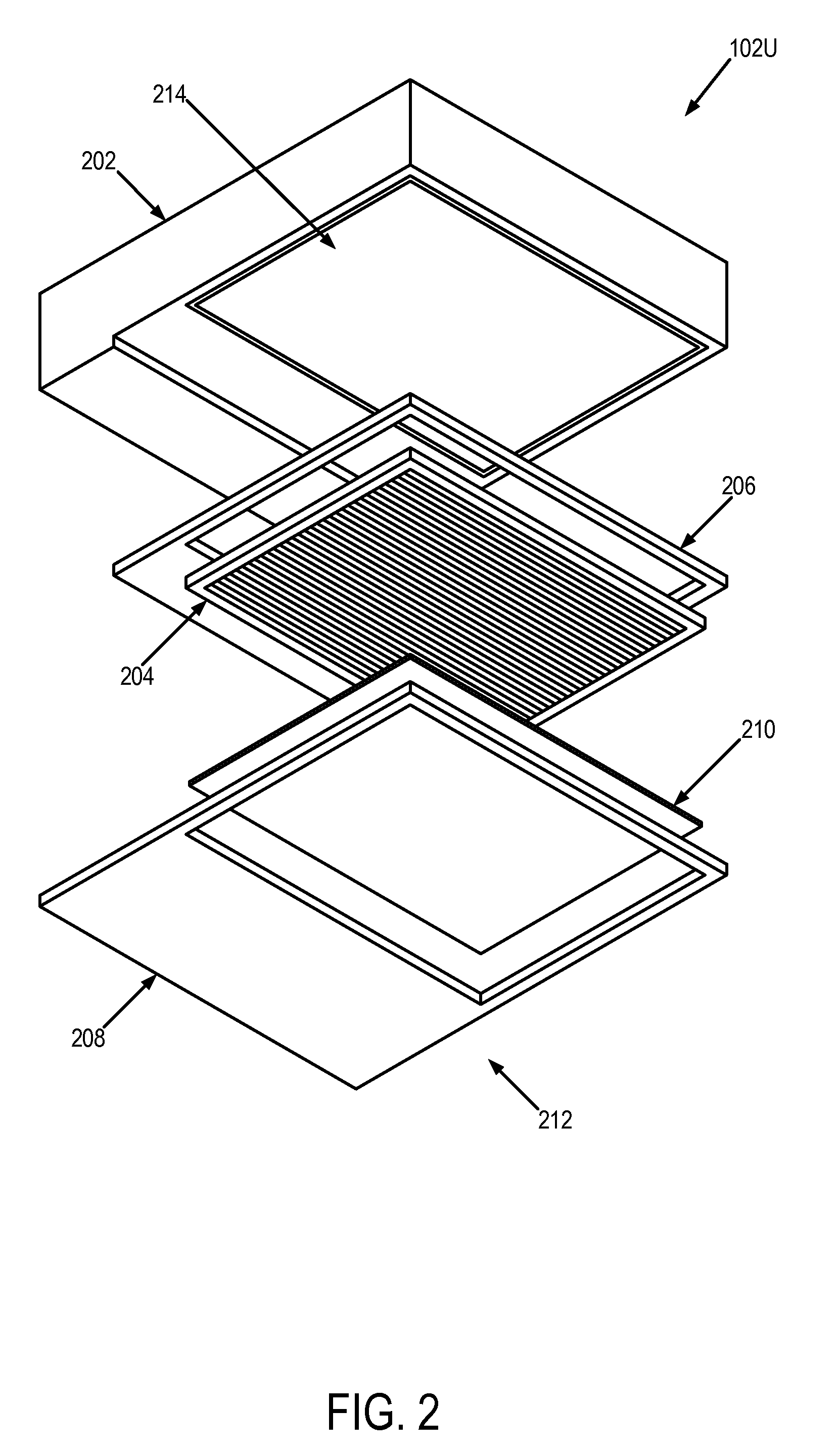
[0013]It is an aspect of the invention to provide an MBI system that includes a support structure arranged proximate to an examination region, a first detector head coupled to the support structure to extend along a first portion of the examination region, and a second detector head coupled to the support structure to extend along a second portion of the examination region. The first detector head includes a first gamma ray detector supported by the support structure and extending to define a first imaging plane proximate to the examination region, and a first collimator extending substantially coplanar with the first imaging plane and arranged between examination region and the first detector. Likewise, the second detector head includes a second gamma ray detector supported by the support structure and extending to define a second imaging plane proximate to the examination region, and a second collimator extending substantially coplanar with the second imaging plane and arranged between examination region and the second detector. The MBI system further includes an adjustable coupling arranged between the first detector head and the support structure. The adjustable coupling permits movement of the first detector head between a first position, in which the first detector head is substantially directly opposed to the second detector head, and a second position, in which the first detector head is not substantially directly opposed to the second detector head.

Problems solved by technology

Despite the success of screening mammography, however, it is also recognized that mammography is a less than perfect screening method.
The limitations of mammography are particularly evident in women with mammographically dense breasts.
The reduced sensitivity of mammography with increasing mammographic density is compounded by the fact that increased density is a significant risk factor for breast cancer.
Given that a dense breast pattern occurs more frequently in younger women, this factor significantly diminishes the value of mammography in the screening of young women who have a high familial risk of breast cancer.
A second major limitation to screening mammography is in the evaluation of women at high risk of breast cancer.
Most of these studies have been performed in women with an average age of forty, so part of the explanation for the poor performance of mammography in these studies may be due to the presence of dense breast patterns in a significant percentage of the mammographic images.
However, while supplemental ultrasound screening uncovers more breast cancers, it also substantially increases the risk of a false positive cancer finding and unnecessary biopsy.
Hence, the use of whole-breast ultrasound as a sole identifier of breast malignancies is questionable.
Even in combination with mammography, the two anatomical techniques have significant limitations.
Currently it is not practical to fuse anatomical images from ultrasound systems and functional images from MBI.
Because of the differences in patient orientation between MBI and ultrasound, the shape of the breast tissue is significantly different between the two modalities and, hence, correlation of an anatomical abnormality with a functional abnormality becomes difficult.
Therefore, accurate co-registration of anatomical images from ultrasound and functional information from MBI is not currently possible.
If the patient is not supine, then the WUS system suffers from a loss in the achievable coverage of the breast tissue.
Therefore, while WUS systems provide better coverage in non-supine patient positions than traditional ultrasound imaging, they are still limited in their applicability to combination with imaging modalities that require non-supine patient positions, such as MBI.

Method used


Image

  • Method and apparatus for dual-modality ultrasonic and nuclear emission mammography
  • Method and apparatus for dual-modality ultrasonic and nuclear emission mammography
  • Method and apparatus for dual-modality ultrasonic and nuclear emission mammography

Examples


Embodiment Construction

[0029]Current schemes for radio-guided breast biopsy are limited in their ability to incorporate additional information from x-ray or ultrasound imaging. Additionally, such schemed only provide a mechanism to localize a lesion and to perform biopsy, but do not provide any supplementary information about the lesion. Thus, the schemes developed for positron emission mammography (“PEM”) and breast specific gamma imaging (“BSGI”) only provide a conventional imaging scheme to localize a lesion, and neither of these systems incorporates any innovative features to enhance or accelerate the biopsy process. With typical imaging times for both PEM and BSGI at around ten minutes per view, the biopsy process can become a lengthy procedure depending on how many images need to be acquired to confirm correct placement of the biopsy needle.

[0030]The MBI system of the present invention provides anatomical correlation information, which is very helpful for aiding a clinician in the decision making pr...


PUM


Abstract

A molecular breast imaging (“MBI”} configured for combined MBI and ultrasound imaging or MBI-guided biopsy is provided. In one configuration, the MBI portion of the system includes two opposed gamma ray detectors, while in another configuration only one gamma ray detector is provided. In two detector configurations, a compression plate is provided to make contact with and apply light compression to the subject under examination. One of the gamma ray detectors is configured to be moveable with respect to the other detector such that the gamma ray detector can be moved away from the examination region, thereby allowing access to the breast for an ultrasound imaging system or a breast biopsy device.

Description

CROSS-REFERENCE TO RELATED APPLICATIONS[0001]This application represents the national stage entry of PCT International Application No. PCT / US2011 / 038698 filed Jun. 1, 2011, which claims the benefit of U.S. Provisional Patent Application Ser. No. 61 / 350,644, filed on Jun. 2, 2010, both of which are incorporated herein by reference in their entirety for all purposes.FIELD OF THE INVENTION[0002]The invention relates to systems and methods for breast imaging. More particularly, the invention relates to systems and methods for combined molecular breast imaging and ultrasound imaging.BACKGROUND OF THE INVENTION[0003]Breast cancer screening has been recommended for many decades, particularly in women over the age of fifty. The combination of early detection and improved therapy in the U.S. has resulted in a significant reduction in breast cancer mortality, with similar reductions being observed in other countries. Despite the success of screening mammography, however, it is also recognized...

Claims


Application Information

Patent Timeline
Patent Type & Authority Patents(United States)
IPC IPC(8): G01T1/164A61B6/00A61B6/03A61B6/04A61B8/00A61B8/08A61B19/00G21K1/02
CPCA61B6/502A61B6/4057A61B6/037G21K1/025A61B8/0825A61B6/0414A61B8/4416A61B6/5247A61B6/4258A61B6/4417A61B6/4291A61B19/201A61B8/485A61B90/11
Inventor O'CONNOR, MICHAEL, K.
Owner MAYO FOUND FOR MEDICAL EDUCATION & RES
Why Eureka
Try Eureka