Surgical training aids and methods of fabrication thereof

a training aid and surgical technology, applied in the field of surgical training aids and methods of fabrication, can solve the problems of cadavers not having the proper feel of living human tissues, animal models not being as accurate as human anatomically, and expensive and often limited supply of human cadavers,

Active Publication Date: 2014-10-28
UNIV OF WESTERN ONTARIO
View PDF10 Cites 55 Cited by
  • Summary
  • Abstract
  • Description
  • Claims
  • Application Information

AI Technical Summary

Benefits of technology

[0011]Hydrogel-based surgical training devices not only have the “feel” of real tissues, such as cardiovascular tissue, but by altering several processing and composition parameters, the hydrogels can be adapted to have the correct mechanical properties of the targeted tissues, including anisotropy, (for example, the aorta and saphenous veins) to give a realistic surgical training experience.

Problems solved by technology

There is an obvious limitation to both of these approaches, where animal models might not be as accurate as human anatomically and human cadavers are costly and often in limited supply.
While human cadavers provide the advantages of presenting the correct anatomy, they disadvantageously do not possess the proper feel of living human tissues, since body is embalmed by submerging in chemicals that change the mechanical properties of the tissues.
Additionally, cadavers are expensive and have disposal issues as they have to be disposed by incineration due their biohazard classification.
An alternative but still costly solution is to provide live animals as surgical teaching resources.
However, live animals do not possess the right physiology, when compared to human tissues and organs.
Furthermore, live animals can necessitate approval of the ethics board and animal rights groups, and also may require expensive animal facilities with well trained veterinary staff.
Unfortunately, such biological samples require special handling, can present problems with transgenic contamination, and require disposal as a ‘biohazard’.
Unfortunately, state-of-the-art anatomic replicates suffer from numerous disadvantages that have hampered their widespread adoption as teaching tools.
Such devices are typically based on silicone rubber, polyurethane, natural rubber, PVC, or foam, and therefore do not replicate properties or the “feel” of the real organs and do not provide an optimal experience to surgical skills training.
While these models may look quite realistic, they fail in producing realistic response in the surgical steps of handling, cutting and suturing, their general ‘feel’ or mechanical behavior is often very different from that of the natural tissues.
Accordingly, such materials do not provide the necessary material properties to be used as effective surgical teaching tools.
The synthetic anastomosis tubes used were shown to significantly underperform animal models with regard to both practicality and accuracy.

Method used


Image

  • Surgical training aids and methods of fabrication thereof
  • Surgical training aids and methods of fabrication thereof
  • Surgical training aids and methods of fabrication thereof

Examples


example 1

Fabrication of Vascular PVA-Hydrogel Surgical Training Devices

[0076]PVA hydrogels that match the mechanical properties of selected cardiovascular tissues, such as coronary arteries, internal mammary artery, saphenous veins, and aorta, were developed.

[0077]PVA (Sigma-Aldrich Canada Co.) with a molecular weight (Mw) of 146,000-186,000, 99+% hydrolyzed was used in all solution preparations. The 10 wt % PVA solutions in distilled water were prepared using a mixed reactor vessel kept at 90° C. for 3 hours under reflux. The PVA concentration can be altered from 5% to 50% to alter final product properties.

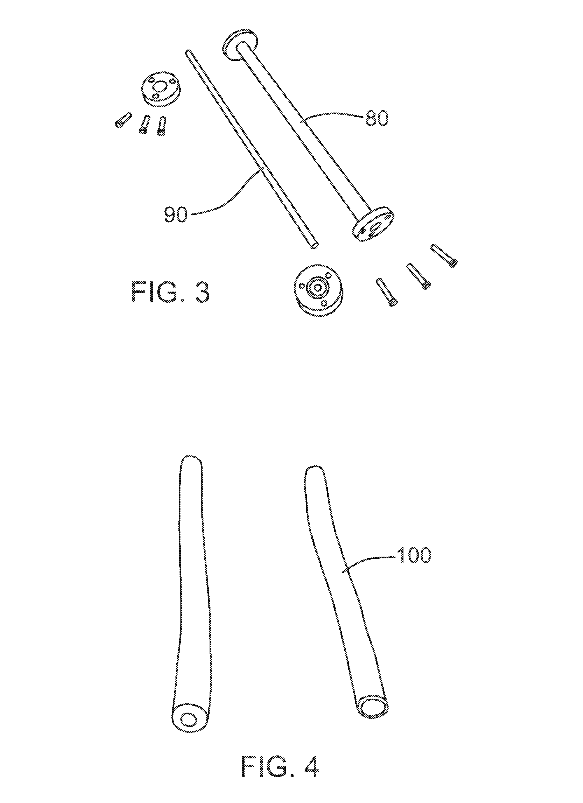
[0078]As shown in FIG. 3, aluminum molds of large diameter 80 (25 mm-aorta) and small diameter 90 (4 mm-veins) conduits were designed and constructed. Six grafts of each diameter (with matching properties of several tissues) were developed and used in surgical training of bypass surgery to test handling, mechanical strength, feel, and suturability. In order to match some of the tissues, d...

example 2

Evaluation of PVA-Hydrogel Vascular Device for Surgical Training

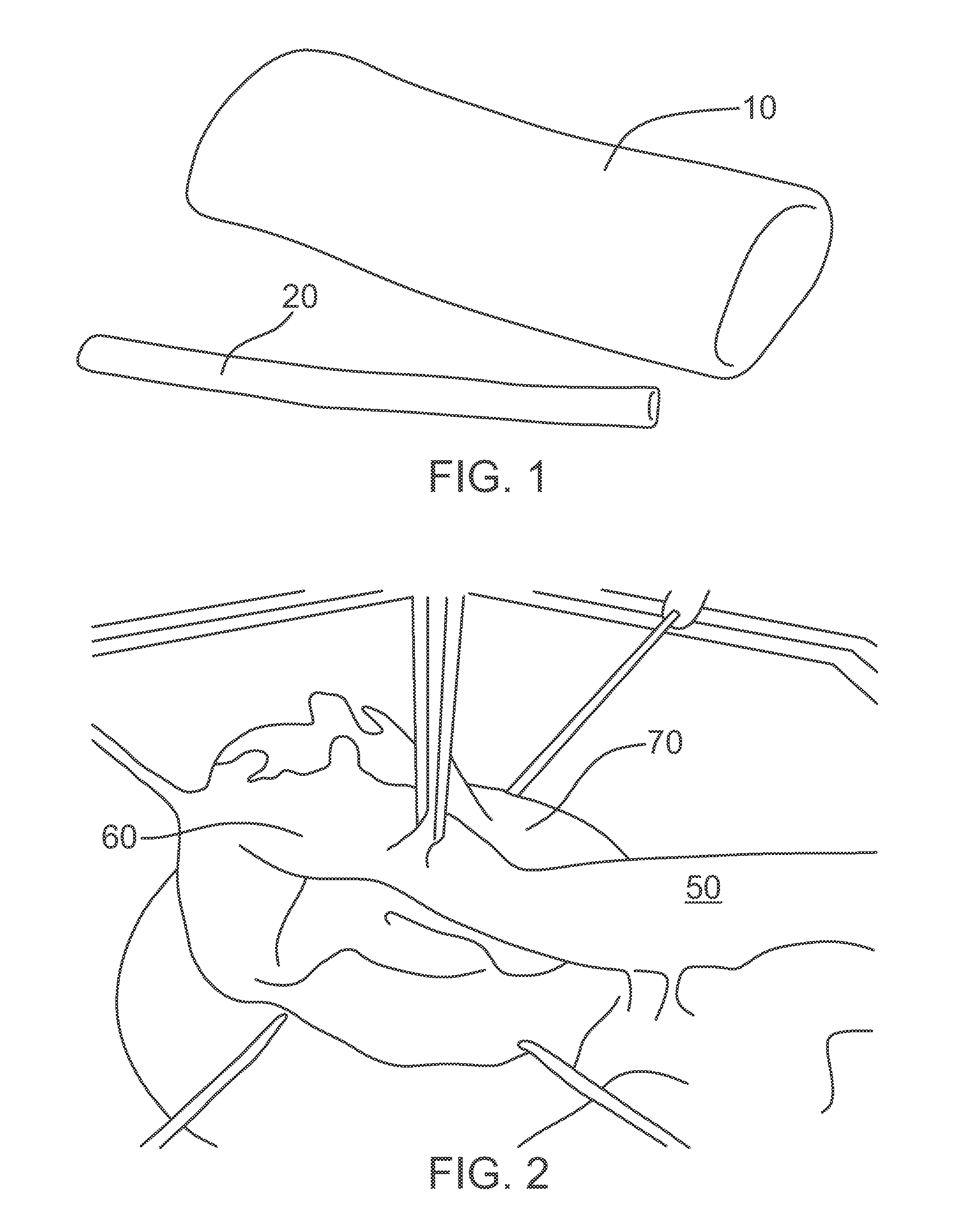
[0081]The 4 mm and the 25 mm diameter PVA-hydrogel conduits were sutured (anastomosed) together to simulate bypass grafting. This exercise was performed by an experienced surgeon. FIG. 5 shows the step-by-step anastomosis of a match of PVA vein 150 to aorta 160.

example 3

Kit for Coronary Artery Bypass Grafting (CABG) Surgical Training

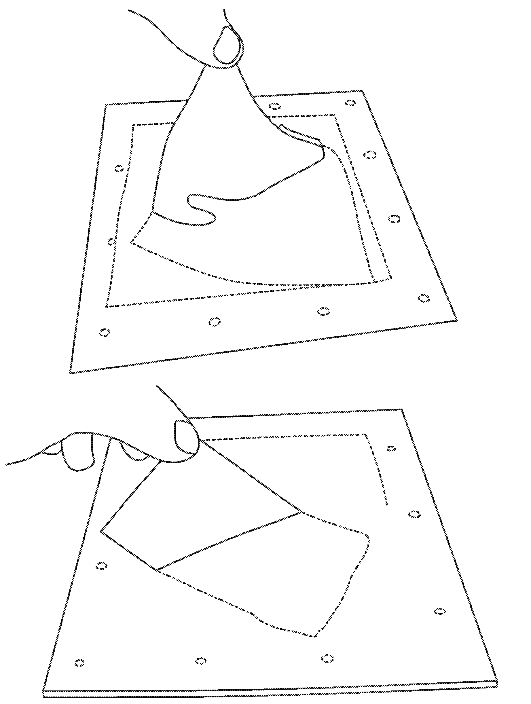
[0082]Another model was developed to provide a PVA-hydrogel surgical training aid that imitates coronary arteries laying on the surface of a synthetic ‘heart’. A mold, comprising a base 200, cover plate 210, and rods 220, was designed to make a block of PVA with 4 parallels arteries protruding from the surface. The arteries ranged in diameters from 1.75, 2, 2.25 and 2.5 mm. The wall thickness was kept constant at 0.5 mm. The mold was filled with PVA solution (processing described before) and cycled up to 6 times to obtain the desired properties, close to myocardium. FIG. 6 shows the mold designed for coronary arteries.

[0083]A surgeon practiced an anastomosis procedure of small diameter PVA conduits (vein) onto the PVA “coronary arteries” on the surface of the surgical aid. This procedure, together with the previous example, imitates the full procedure implemented in CABG surgery, where the bypass graft is anastomosed to...


PUM

PropertyMeasurementUnit
diameteraaaaaaaaaa
diameteraaaaaaaaaa
thicknessaaaaaaaaaa

Abstract

The present invention provides surgical training aids formed from hydrogels and adapted to exhibit realistic mechanical properties mimicking those of real organs. Surgical training aids are preferably fabricated by subjecting a concentration of polyvinyl alcohol to freeze-thaw cycles in a mold designed to approximate the shape of an organ, and process parameters are selected to tailor the mechanical properties of the formed hydrogel to those of the organ simulated by the surgical aid. The mechanical properties of the hydrogel forming the surgical training aid may be tailored by incorporating bacterial cellulose and by applying strain during hydrogel formation, thereby producing controlled anisotropy.

Description

CROSS-REFERENCE TO RELATED APPLICATION[0001]This application is a National Phase application claiming the benefit of PCT / CA2010 / 001453 filed on Sep. 22, 2010, in English, which further claims priority to U.S. Provisional Application No. 61 / 244,690 titled “SURGICAL TRAINING AIDS AND METHODS OF FABRICATION THEREOF” and filed on Sep. 22, 2009, the entire contents of which are incorporated herein by reference.FIELD OF THE INVENTION[0002]This invention relates to the field of surgical skills training, and more particularly relates to the field of surgical models, simulators, aids, kits and methods of training for surgical procedures involving anastomosis of tubular structures, as well as dissecting, cutting and suturing of soft tissues and organ models.BACKGROUND OF THE INVENTION[0003]Traditionally, surgical training and skill development has been performed in the operating room on living patients. However, there are increasing medical, legal and ethical concerns about the use of patient...

Claims


Application Information

Patent Timeline
Patent Type & Authority Patents(United States)
IPC IPC(8): G09B23/30B29C39/42B29L31/00C08J3/075B29K105/00B29C39/02B29C39/00G09B23/28B29K29/00
CPCB29L2031/7532G09B23/306C08J3/075G09B23/28C08J2329/04B29C39/003B29K2029/04B29C39/42B29C39/025B29K2105/0061
Inventor MILLON, LEONARDOWAN, WANKEIQUANTZ, MACKENZIE
Owner UNIV OF WESTERN ONTARIO
Why Eureka
Try Eureka