
Method for computing pharmacokinetic parameters in MRI

a pharmacokinetic parameter and mri technology, applied in the field of enhancing the accuracy of pharmacokinetic parameter computation in the mri system, can solve the problems of insufficient reliability of tissue classification, inability to determine the threshold of normal, benign, and malignant tissues, and insufficiently reliable tissue classification to provide a conclusive diagnosis. , to achieve the effect of improving the visibility of internal body structures

Inactive Publication Date: 2015-04-21
RAJIV GANDHI CANCER INST & RES CENT
View PDF3 Cites 3 Cited by
  • Summary
  • Abstract
  • Description
  • Claims
  • Application Information

AI Technical Summary

Benefits of technology

The patent aims to improve the accuracy of tissue classification using magnetic resonance imaging (MRI) techniques. It proposes using phantoms for normalization of the contrast agent uptake time constant (T10) and pharmacokinetic parameters to enhance the accuracy of computed T1 values in each data point. This technique can be applied to any MRI system used for tissue classification. Contrast agents used for enhancing visibility of internal body structures include Gadolinium and other FDA-approved agents, as well as supermagnetic iron oxide compounds. Overall, this patent provides a method to improve the accuracy of identifying malignant, benign, and normal tissues using MRI.

Problems solved by technology

However, to date conventional MRI has not been capable enough to distinguish accurately between normal, benign, and malignant tissues.
This is primarily because tissues have a number of distinguishing characteristics, which change for each patient and the tissue being monitored, and therefore a fixed threshold for classifying the tissue is not possible.
However, the results of classification of tissues are currently insufficiently reliable to provide a conclusive diagnosis.
Also, the intrinsic T1 value varies with the machine parameters, resulting in incorrect computation of net T1 value when the contrast is given, which as a result leads to inaccurate computation of pharmacokinetic parameters that are used for tissue classification.
Also, if a particular voxel is showing a high enhancement, it is difficult to conclude that whether this is due to the uptake of contrast agent or the intrinsic T1 value of the tissue.
Thus, one cannot predict whether it is a physiologically-based effect (high uptake of contrast agent) or an intrinsic effect (because of the T1 value of that type of tissue).

Method used


Image

  • Method for computing pharmacokinetic parameters in MRI
  • Method for computing pharmacokinetic parameters in MRI
  • Method for computing pharmacokinetic parameters in MRI

Examples


Embodiment Construction

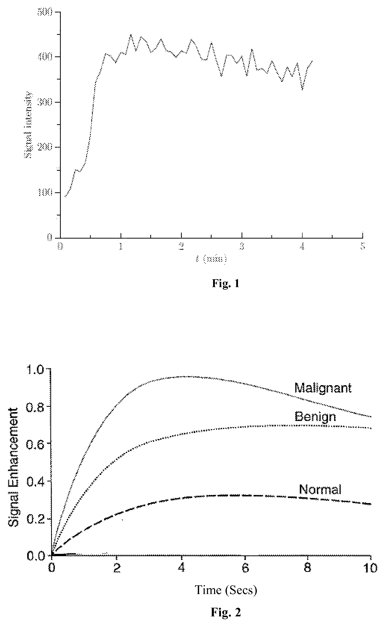
[0028]The present invention relates to an effective dynamic contrast enhanced magnetic resonance imaging (DCE-MRI) of tissues such as breast cancer tissues. The present invention further provides a method of enhancing and improving accuracy of calculated pharmacokinetic parameters such as Ktrans for effective detection and differentiation of malignant, benign and normal tissues.

[0029]In an embodiment, a contrast agent / tracer is injected into the body of the patient and a time sequence of MRI volumes is acquired. Such a time sequence image could be acquired every 2 seconds or 4 seconds, which increases the number of data points being plotted. Use of contrast agents significantly enhances T1-weighted MRI readings. Contrast agents are transported throughout the body by vascular systems (arteries, arterioles, capillaries, veins and other types of vessels), in which process, the contrast diffuses across the vessel walls into the surrounding tissue which comprises tissue cells and extrace...


PUM


Abstract

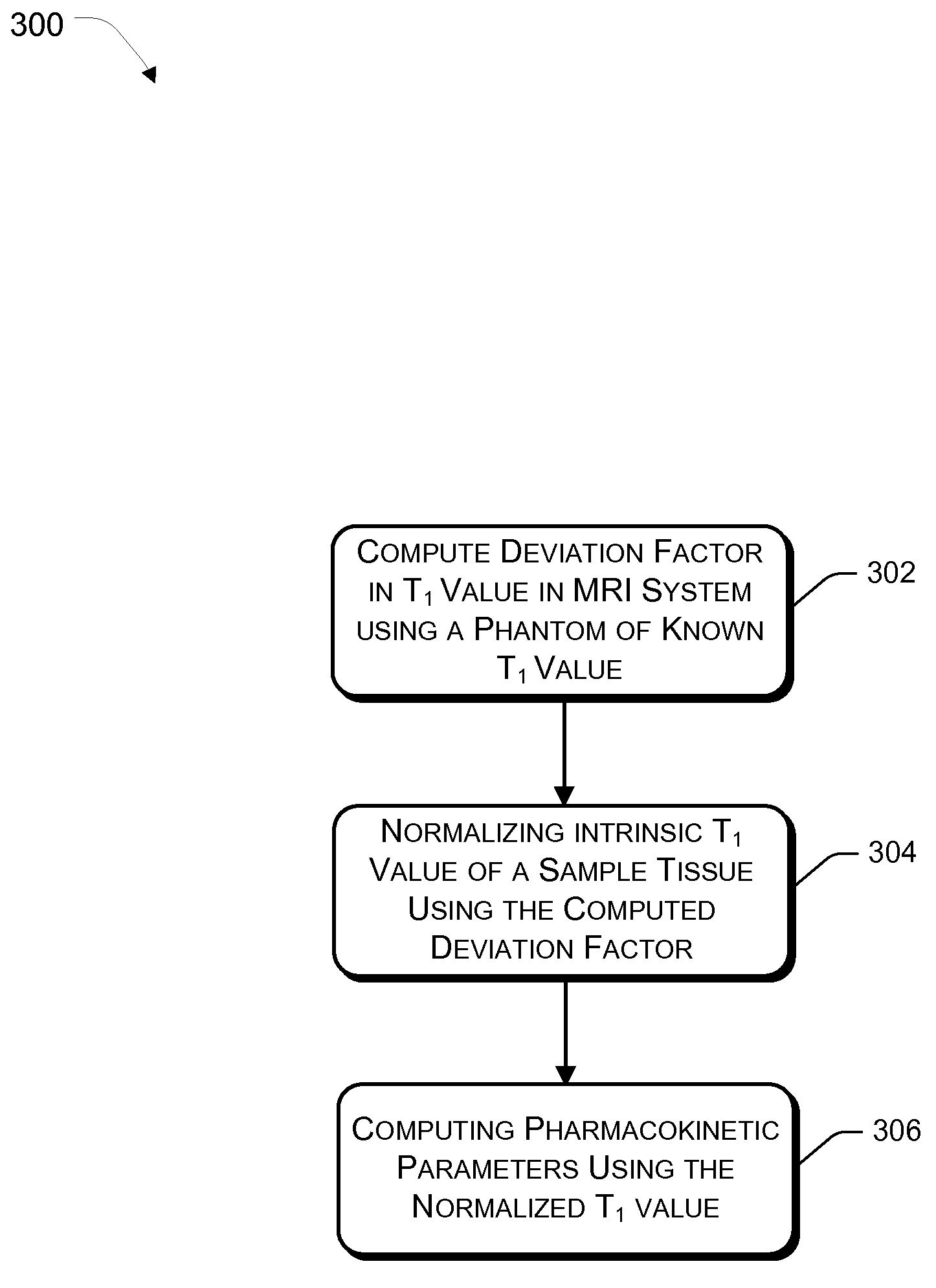
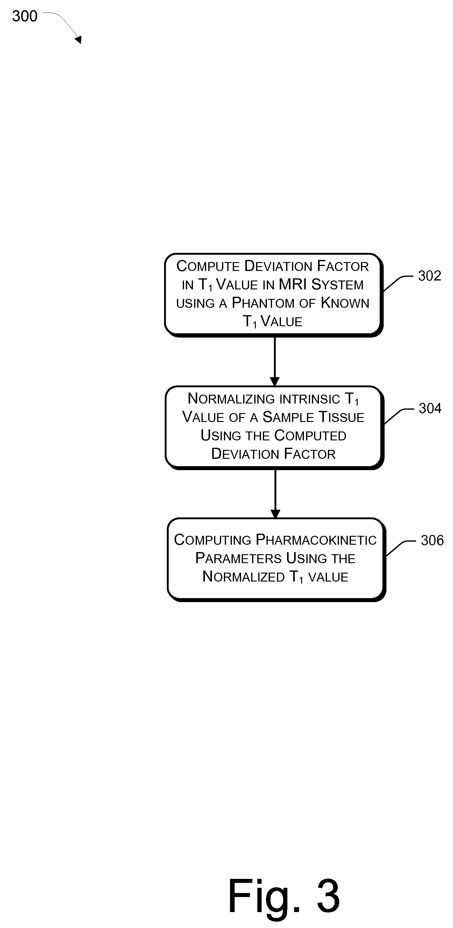
The present invention relates to a method of dynamic contrast magnetic resonance imaging aimed to improve characterization of tissue image by improving accuracy of computed pharmacokinetic parameters such as Ktrans (transfer constant across capillary membrane; a pharmacokinetic parameter of tissue perfusion) using T1W Fast DCE-MRI technique to distinguish between malignant, benign and normal tissues. A phantom and a contrast agent are used in the proposed MRI system for computing accurate T1 value of a tissue after the contrast is injected based on an intrinsic T10 value the tissue, wherein the intrinsic T10 value is adjusted and / or normalized to improve accuracy of the T1 value computed, which in turn is used for computation of pharmacokinetic parameters at least one of Ktrans.

Description

FIELD OF INVENTION[0001]The present subject matter relates to a method for enhancing accuracy of pharmacokinetic parameter computation in MRI system. More specifically, the present subject matter relates to a method for enhancing accuracy of computation of pharmacokinetic parameters such as Ktrans, Kep, and Ve in MRI for more accurate, reliable, and reproducible evaluation and classification of tissues.BACKGROUND OF THE INVENTION[0002]Magnetic resonance imaging (“MRI”) is a well-known, highly useful technique for diagnosing abnormalities in biological tissues. MRI can detect abnormalities that are difficult or impossible to detect by other techniques, without the use of x-rays or invasive procedures.[0003]However, to date conventional MRI has not been capable enough to distinguish accurately between normal, benign, and malignant tissues. This is primarily because tissues have a number of distinguishing characteristics, which change for each patient and the tissue being monitored, an...

Claims


Application Information

Patent Timeline
Patent Type & Authority Patents(United States)
IPC IPC(8): G01V3/00G01R33/48G01R33/50G01R33/56G01R33/563
CPCG01R33/50G01R33/56366G01R33/4828G01R33/5601
Inventor JENA, AMARNATHMEHTA, SHASHI BHUSHANTANEJA, SANGEETASARKAR, URMI
Owner RAJIV GANDHI CANCER INST & RES CENT
Learn More