System and method for dual energy and/or contrast enhanced breast imaging for screening, diagnosis and biopsy

a breast imaging and contrast enhancement technology, applied in the field of medical imaging, can solve the problems of difficult to determine whether a detected abnormality is associated with a cancerous or benign lesion, long procedure time, and confusion in image interpretation and diagnosis, so as to facilitate x-ray screening and diagnosis of patients, facilitate identification of a position, and facilitate the effect of screening and diagnosis

Active Publication Date: 2015-04-28
HOLOGIC INC
View PDF16 Cites 33 Cited by
  • Summary
  • Abstract
  • Description
  • Claims
  • Application Information

AI Technical Summary

Benefits of technology

[0013]The patent specification describes x-ray imaging systems and methods that facilitate x-ray screening and diagnosis of patients, particularly of patients' breasts, and particularly for abnormalities characterized by suspicious vascularity. In a non-limiting example, combination of imaging modes are used, preferably in a single breast compression, to obtain a collection of x-ray images that provide unexpectedly better facility of screening and diagnosis of such abnormalities. As one example, the new system and method are used to image a patient's breast after an x-ray contrast agent has been introduced in the breast. A selected time after injecting the contrast agent, the system obtains 3D slice images representing respective slices of a patient's breast. These 3D slice images are formed by computer-processing, through a reconstruction algorithm, a multiplicity of x-ray 2D tomosynthesis projection images of the breast taken at respective angles of an imaging x-ray beam to the breast. The system also obtains a 2D combination image of a low-energy 2D x-ray mammogram and a high-energy 2D x-ray mammogram of the breast. These 2D projection images and low-energy and high-energy mammograms preferably are obtained in a single compression of the patient's breast. The system displays, preferably concurrently, the 2D combination image and one or more of the 3D slice images. The displayed combination 2D image facilitates identification of a position of a possible vascular abnormality in two dimensions, and the 3D slice images facilitate identification of the position of the abnormality in three dimensions and enables visualization of the appearance of the abnormality in respective slice images.

Problems solved by technology

While screening x-ray mammography is recognized as the most effective method for early detection of breast cancer, it also presents challenges in that in some cases it may be difficult to determine whether a detected abnormality is associated with a cancerous or benign lesion.
One reason for this is that a mammogram Mp is a two dimensional projection image representating a three dimensional structure, and overlapping structures in the compressed breast may confound image interpretation and diagnosis.
Although contrast enhanced breast MRI (CEMRI) can be effective in imaging breast cancer it has limitations including high cost, long procedure time, enhancement of benign abnormalities such as fibroadenomas, and inability to image women with metal clips or claustrophobia.

Method used


Image

  • System and method for dual energy and/or contrast enhanced breast imaging for screening, diagnosis and biopsy
  • System and method for dual energy and/or contrast enhanced breast imaging for screening, diagnosis and biopsy
  • System and method for dual energy and/or contrast enhanced breast imaging for screening, diagnosis and biopsy

Examples


Embodiment Construction

[0027]The examples of systems and methods described in this patent specification leverage and combine advantages of one or more image acquisition modes, including two-dimensional (2D), three-dimensional (3D), dual-energy (DE) and contrast-enhancement (CE) imaging to provide a breast imaging system with improved sensitivity and specificity and with benefits for more efficacious screening and diagnosis, greater convenience for the radiologist and better patient workflow.

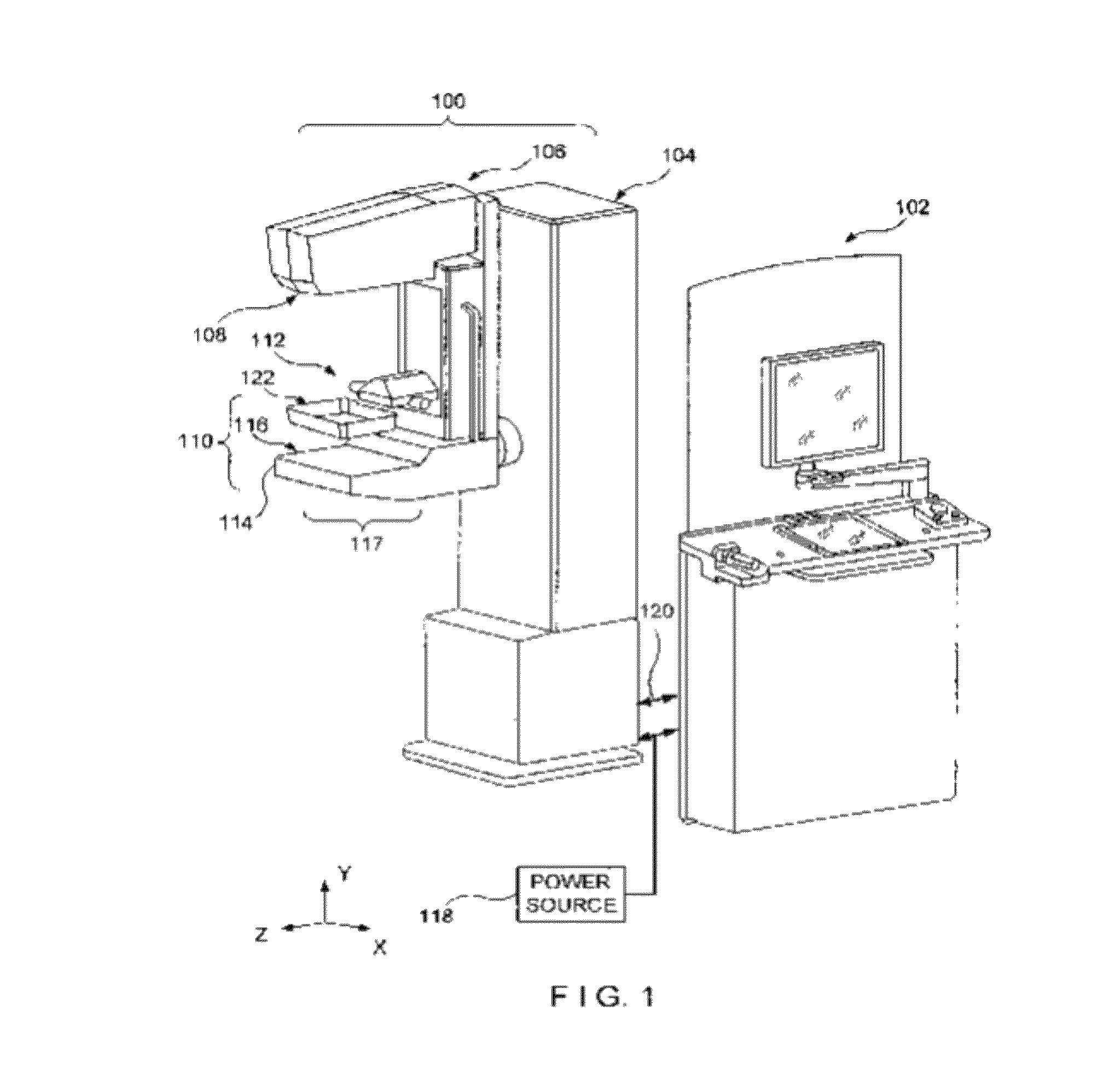
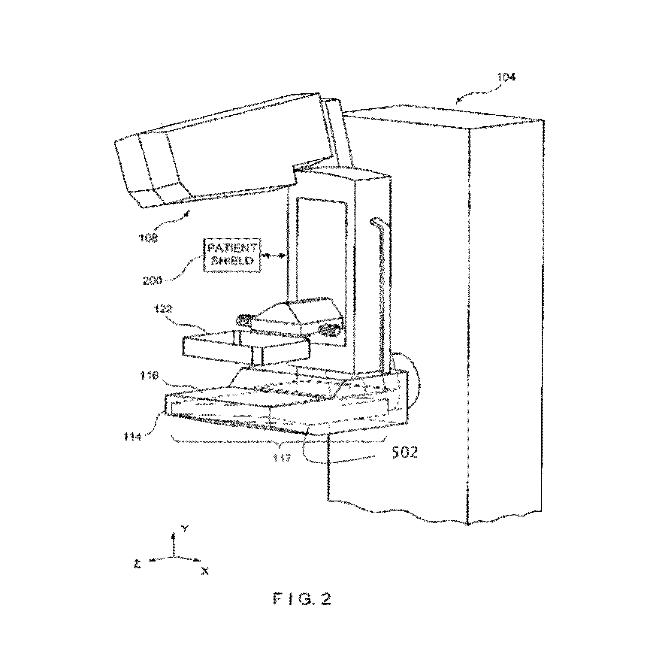
[0028]A system according to one example includes an x-ray source including one or more x-ray filters, an imaging x-ray detector, and an immobilization mechanism positioned between the x-ray source and the detector for immobilizing an object to be imaged such as a patient's breast. During image acquisition, X-rays of two or more different energy ranges are generated from the x-ray source by varying at least one x-ray source acquisition parameter, including but not limited to the x-ray filters and x-ray kV. The x-rays pr...


PUM


Abstract

Systems and methods for x-ray imaging a patient's breast in combinations of dual-energy, single-energy, mammography and tomosynthesis modes that facilitate screening for and diagnosis of breast abnormalities, particularly breast abnormalities characterized by abnormal vascularity.

Description

REFERENCE TO RELATED APPLICATION[0001]This patent application claims the benefits of provisional application No. 61 / 450,304 filed Mar. 8, 2011 and incorporates by reference the specification and drawings thereof.FIELD[0002]This patent specification relates to medical imaging and more specifically to a system that enables selection among a plurality of different imaging modes, a plurality of different imaging processes, image acquisition parameters and image processing techniques.BACKGROUND[0003]Several references are listed at the end of the disclosure portions of this patent specification and are referred to below by numbers in parenthesis. These references as well as prior patents identified in this patent specification are hereby incorporated by reference.[0004]In the U.S. breast cancer mortality is second only to that of lung cancer for women. Because of its role in early tumor detection, x-ray mammography has become the most commonly used tool for breast cancer screening, diagn...

Claims


Application Information

Patent Timeline
Patent Type & Authority Patents(United States)
IPC IPC(8): A61B5/05A61B6/00A61B6/02A61B6/12
CPCA61B6/025A61B6/4417A61B6/463A61B6/466A61B6/502A61B6/5235A61B6/482A61B6/12A61B6/4035A61B6/481F04C2270/041A61B10/04
Inventor SMITH, ANDREW P.STEINJING, ZHENXUEREN, BAORUINIKLASON, LORENRUTH, CHRISTOPHERSHAW, IAN
Owner HOLOGIC INC
Why Eureka
Try Eureka