Method for delineation of tissue lesions

a tissue lesions and computer assisted technology, applied in image data processing, instruments, character and pattern recognition, etc., can solve the problems of lack of established thresholds, inability to reliably use automated image processing methods, time-consuming and costly manual determination of dwi/pwi mismatch, etc., to improve speed and/or reproducibility, reduce user intervention, the effect of fast estimation

Active Publication Date: 2015-05-19
REGION MIDTJYLLAND +1
View PDF9 Cites 0 Cited by
  • Summary
  • Abstract
  • Description
  • Claims
  • Application Information

AI Technical Summary

Benefits of technology

[0010]Furthermore, it may be seen as an advantage of the invention that it may yield reproducible results. Another advantage may be that the method may be implemented so as to operate in a fully automated manner.
[0011]It is a further object of the present invention to provide an alternative to the prior art.
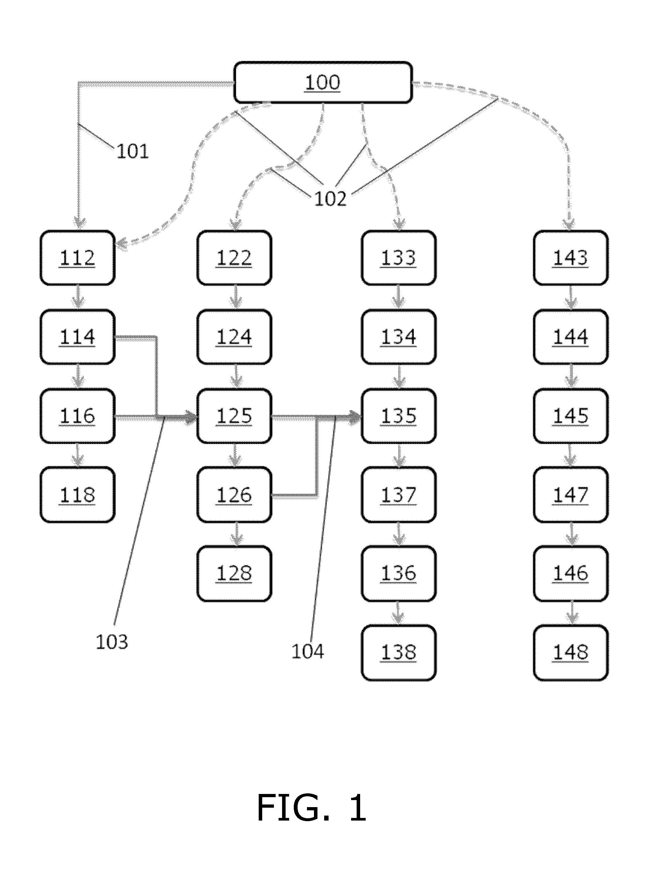
[0012]Thus, the above described object and several other objects are intended to be obtained in a first aspect of the invention by providing a method for estimating a measure of a penumbra size of a penumbra in biological tissue, the method comprising the steps of
[0013]receiving a first image, the first image comprising data representative of a spatially resolved perfusion parameter,
[0014]receiving a second image, the second image comprising data representative of a spatially resolved diffusion parameter,
[0015]identifying a first area on the first image, which first area corresponds to a perfusion lesion,

Problems solved by technology

Such manual determination of DWI / PWI mismatch can be time consuming and costly.
Such automated image processing methods may not be reliable since they may not be able to distinguish between a lesion volume and a volume outside of the lesion.
This approach, however, suffers from the lack of established thresholds, such as perfusion thresholds, applicable across individuals.

Method used


Image

  • Method for delineation of tissue lesions
  • Method for delineation of tissue lesions
  • Method for delineation of tissue lesions

Examples


example 1

Segmentation of an Image Comprising TTP Data

Materials & Methods

Morphological Grayscale Reconstruction

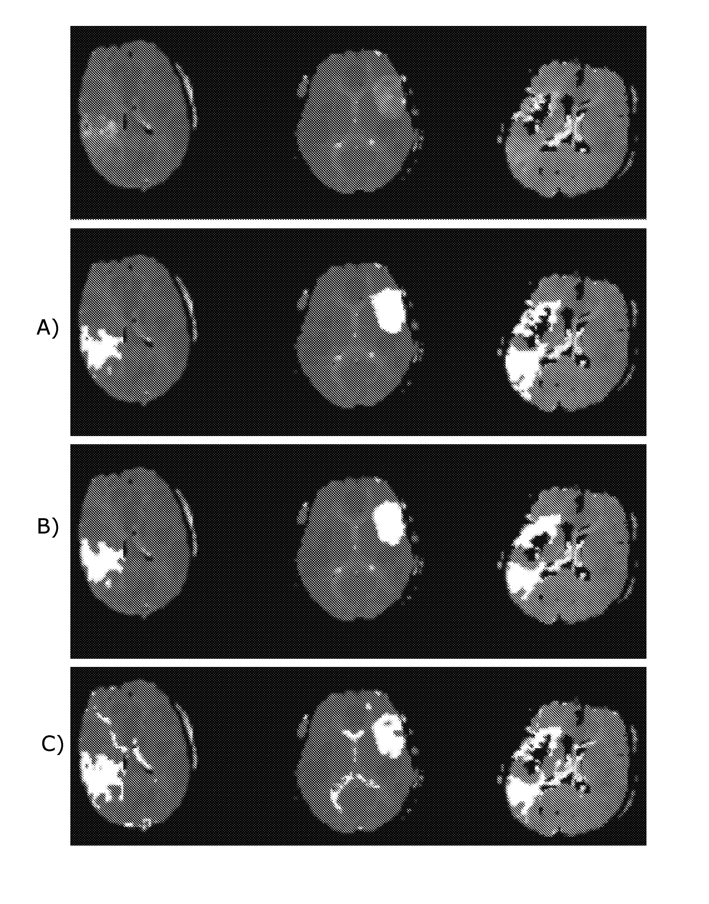
[0138]Finding a voxel within perfusion lesions, so-called seed point, appear easily discernible by eye, however, searching for the maximum of the image intensities often leads to a false positive seed point e.g. high intensities in regions of the image corresponding to irrelevant regions, such as eyes, cerebrospinal fluid (CSF), intensities outside the brain etc. To automatically detect a seed point, we use morphological grayscale reconstruction to truncate small spikes while preserving the edges of TTP lesion and homogenize the background of TTP maps. Compared with conventionally smoothing e.g. with a Gaussian kernel, morphological grayscale reconstruction is less sensitive to spikes and for that reason more suitable for seed point detection. In the present context, spikes are understood to be areas which are relatively small compared to a lesion, and which comprise data points with...

example 2

Segmentation of an Image of Type MTT Data

Materials and Methods

Level Sets

[0163]The approach emerges from the assumption that the average MTT value, Mhypo, in a hypoperfused region is higher than the average normal MTT value, Mnorm. In the hypoperfused area, MTT values will vary around Mhypo, while in normal tissue they are close to Mnorm. This corresponds to low mean squared errors between image MTT values, MTT(x,y), and the respective averages. Identifying the lesion, such as the ischemic lesion, can then be formulated as finding a smooth, closed curve C, which minimizes the total variation (Eq. (1))

E=∫Inside C(MTT(x,y)−Mhypo)2dxdy+∫Outside C(MTT(x,y)−Mnorm)2dxdy

[0164]Since ischemic regions are not necessarily coherent, for instance in cases with occlusions causing lesions in the watershed areas, it is important that C can represent multiple curves. This can be ensured by defining C implicitly as the zero-level set of a function φ which assigns a value to each point the plane, C={φ...

example 3

Segmentation of an Image Obtained by DWI

Materials & Methods

Morphological Grayscale Reconstruction

[0190]DWI lesion appears easily discernible by eye as a hyperintense region, although the diffusion lesion contains artefactual hypointensities and hyperintensities are present throughout the image due to anatomy and noise artifacts. Therefore simple thresholding of the image intensities leads to both high false positive and false negative rates as indicated in FIG. 11B. Typical image blurring, such as convolution by Gaussian kernel, remedies artifacts to some extent, although at the potential cost of ‘moving’ the lesion boundary. Morphological grayscale reconstruction uniquely truncates high peaks in the image while tending to preserve original edges of coherent areas, as determined by the kernel size. We use morphological grayscale reconstruction to enhance the contrast between the DWI lesion and the background. Compared with conventional blurring filter, morphological grayscale recons...


PUM


Abstract

The present invention relates to a method for estimating a measure of a penumbra size of a penumbra in biological tissue wherein first and second images obtained respectively by perfusion weighted imaging (PWI) and diffusion weighted imaging (DWI) are analyzed, and wherein the analysis of the first image includes application of a level set method, and the analysis of the second image includes application of a grayscale morphological operation. In a further embodiment of the invention, a connected component labeling algorithm maybe applied on any one of the first and second image. The invention further relates to a system, a computer program product and use of a corresponding method.

Description

CROSS REFERENCE TO RELATED APPLICATIONS[0001]This application is a U.S. National Phase Application of PCT International Application Number PCT / DK2011 / 050493, filed on Dec. 16, 2011, designating the United States of America and published in the English language, which is an International Application of and claims the benefit of priority to Danish Patent Application No. PA 2010 70558, filed on Dec. 17, 2010, and Danish Patent Application No. PA 2011 70070, filed on Feb. 7, 2011. The disclosures of the above-referenced applications are hereby expressly incorporated by reference in their entireties.FIELD OF THE INVENTION[0002]The present invention relates to a method of computer assisted delineation of tissue lesions, in particular the invention relates to computer assisted delineation of tissue lesions in the brain of a stroke patient. The invention further relates to a system, a computer program product and use of a corresponding method.BACKGROUND OF THE INVENTION[0003]In the case of ...

Claims


Application Information

Patent Timeline
Patent Type & Authority Patents(United States)
IPC IPC(8): G06K9/00G06T11/00G06T7/00
CPCG06T7/0012G06T2207/30016G06T7/0089G06T2207/10088G06T11/003G06T2207/20036G06T7/0091G06T7/149G06T7/155G06T1/00G06T7/00
Inventor NAGENTHIRAJA, KARTHEEBANMOURIDSEN, KIMRIBE, LARS RIISGAARD
Owner REGION MIDTJYLLAND
Why Eureka
Try Eureka