
Methods and systems for segmentation in echocardiography

a technology of echocardiography and methods, applied in the field of methods and systems for segmentation in echocardiography, can solve the problems of difficult automation of the workflow for septum thickness measurement, increased complexity of visualization and analysis of acquired data, and variability between and intra-observer

Active Publication Date: 2015-09-29
GENERAL ELECTRIC CO
View PDF8 Cites 4 Cited by
  • Summary
  • Abstract
  • Description
  • Claims
  • Application Information

AI Technical Summary

Benefits of technology

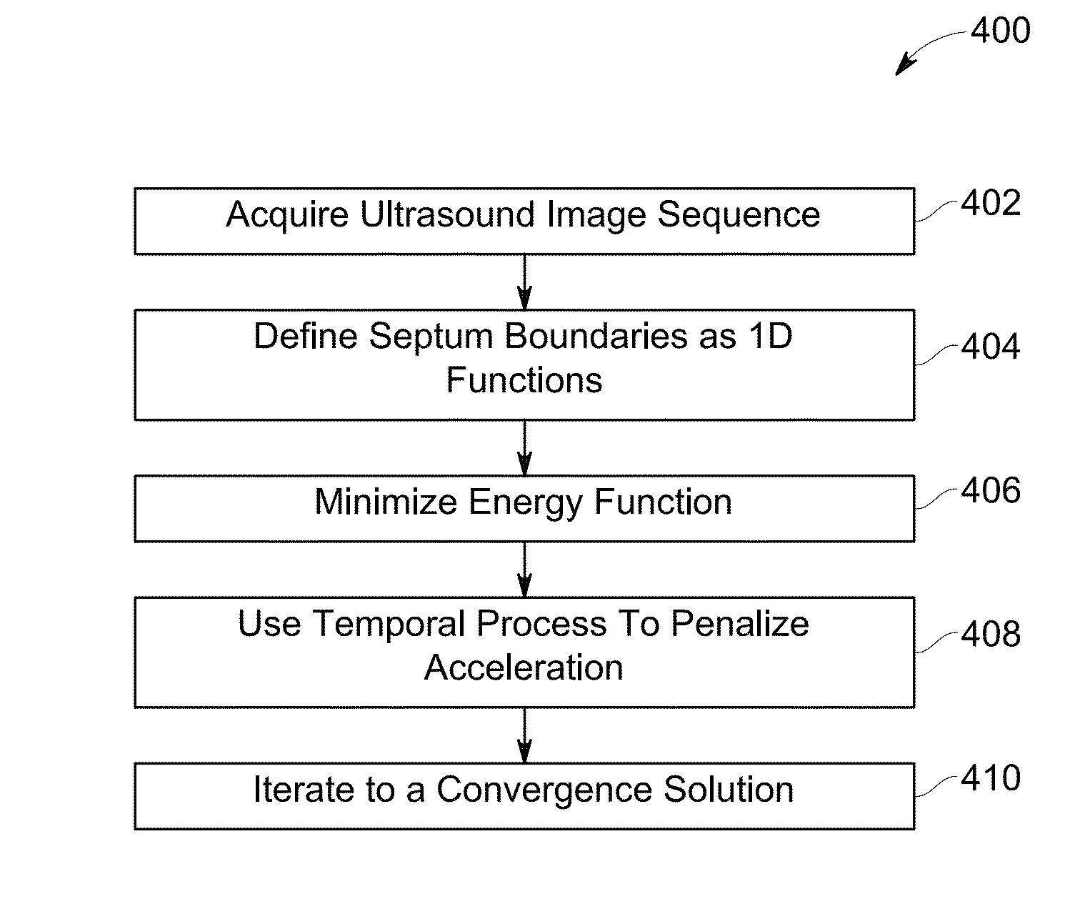
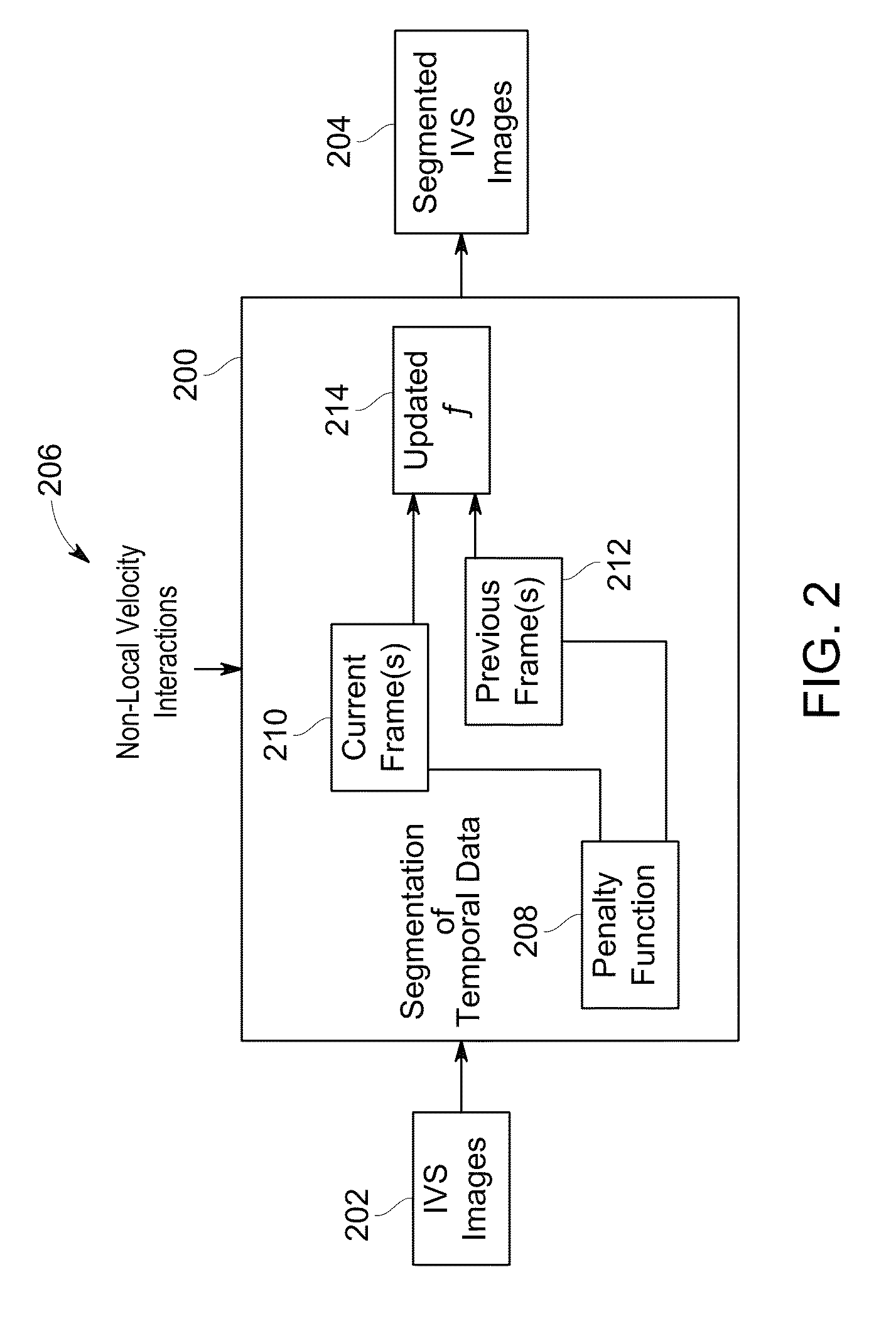
The patent text describes a method and system for automatically segmenting a cardiac structure using echocardiographic images. This is done by defining a search space using 1D profiles and using an energy-based function constrained by non-local temporal priors to automatically segment the contour of the cardiac structure. The result is a more efficient and accurate process for identifying and measuring cardiac structures using ultrasound technology. This can be useful for diagnostic and therapeutic applications in cardiovascular medicine.

Problems solved by technology

Compared to conventional 2D echocardiography, 4D echocardiography increases the complexity of visualization and analysis of the acquired data.
However, because the measurements are performed manually, inter- and intra-observer variability occurs as a result of observer variability based on experience and expertise.
Automation of the workflow for septum thickness measurement is challenging and known methods may not perform satisfactorily for use in real time.
Within the workflow, identifying the septum border is an important and challenging first step.
In particular, noise and in-homogeneities induced by near field haze represent challenges for achieving a good segmentation result.
These challenges result in the need for algorithms that advance the initialization closer to the septum boundary.
However, in some cases, such as where the septum boundary has low contrast resulting in large boundary gaps, the known segmentation methods fail or fail to perform satisfactorily.
Moreover, other approaches, such as region based active contour approaches, also suffer from drawbacks, such as the infeasibility of generating shape atlases for the septum given the large inter-patient shape variability and non-rigid deformation across frames, as well as failure of the segmentation when constraining the width where the septum boundary has low contrast.

Method used


Image

  • Methods and systems for segmentation in echocardiography
  • Methods and systems for segmentation in echocardiography
  • Methods and systems for segmentation in echocardiography

Examples


Embodiment Construction

[0023]The following detailed description of certain embodiments will be better understood when read in conjunction with the appended drawings. To the extent that the figures illustrate diagrams of the functional blocks of various embodiments, the functional blocks are not necessarily indicative of the division between hardware circuitry. Thus, for example, one or more of the functional blocks (e.g., processors or memories) may be implemented in a single piece of hardware (e.g., a general purpose signal processor or random access memory, hard disk, or the like). Similarly, the programs may be stand alone programs, may be incorporated as subroutines in an operating system, may be functions in an installed software package, and the like. It should be understood that the various embodiments are not limited to the arrangements and instrumentality shown in the drawings.

[0024]As used herein, an element or step recited in the singular and proceeded with the word “a” or “an” should be unders...


PUM


Abstract

Methods and systems for segmentation in echocardiography are provided. One method includes obtaining echocardiographic images and defining a search space within the echocardiographic images using a pair of one-dimensional (1D) profiles. The method also includes using an energy based function constrained by non-local temporal priors within the defined search space to automatically segment a contour of a cardiac structure with the 1D profiles.

Description

CROSS REFERENCE TO RELATED APPLICATIONS[0001]This application claims priority to and the benefit of the filing date of U.S. Provisional Application No. 61 / 766,596, filed on Feb. 19, 2013, entitled “Methods and Systems for Segmentation in Echocardiography,” which is hereby incorporated by reference in its entirety.BACKGROUND[0002]Different methods for quantitative analysis of three-dimensional (3D) and four-dimensional (4D) echocardiograms are known. With 3D and 4D echocardiography, different image slices may be extracted from acquired volumes, which may be acquired as a series of image frames covering the cardiac cycle.[0003]In 4D echocardiography, for example, a sequence of volumetric images of a patient's heart may be acquired using an ultrasound system. Compared to conventional 2D echocardiography, 4D echocardiography increases the complexity of visualization and analysis of the acquired data. Thus, a high degree of manual interaction often has to be performed to extract clinical...

Claims


Application Information

Patent Timeline
Patent Type & Authority Patents(United States)
IPC IPC(8): G06K9/00G06T7/00
CPCG06T7/0097G06T7/0089G06T7/0012G06T7/0083G06T2207/10016G06T2207/10132G06T2207/20116G06T2207/30048G06T7/12G06T7/149G06T7/174
Inventor THIRUVENKADAM, SHESHADRISUBAMANIAN, NAVNEETHGUPTA, MITHUN DAS
Owner GENERAL ELECTRIC CO
Learn More