System of obtaining the echocardiography by the dummy gullet passing and the method for realizing the same

A technology of echocardiography and its implementation method, which is applied in the field of medical imaging technology-ultrasound medical imaging, which can solve the disadvantages for beginners in learning and mastering echocardiography, the unclear display of anatomical structure of the three-dimensional visualization model of the heart, and the lack of thin-section anatomical contrast and other issues, to achieve the effect of increasing popularization, reducing incidence, and avoiding pain

Inactive Publication Date: 2007-09-19
THE FIRST AFFILIATED HOSPITAL OF THIRD MILITARY MEDICAL UNIVERSITY OF PLA
View PDF0 Cites 24 Cited by
  • Summary
  • Abstract
  • Description
  • Claims
  • Application Information

AI Technical Summary

Problems solved by technology

But there are also some shortcomings: (1) The three-dimensional visualization model of the heart established on the basis of two-dimensional ultrasound sections still has the disadvantage that the anatomical structure is not clearly displayed; (2) the system only displays the cutting position of the probe and its

Method used


Image

  • System of obtaining the echocardiography by the dummy gullet passing and the method for realizing the same

Examples


Embodiment Construction

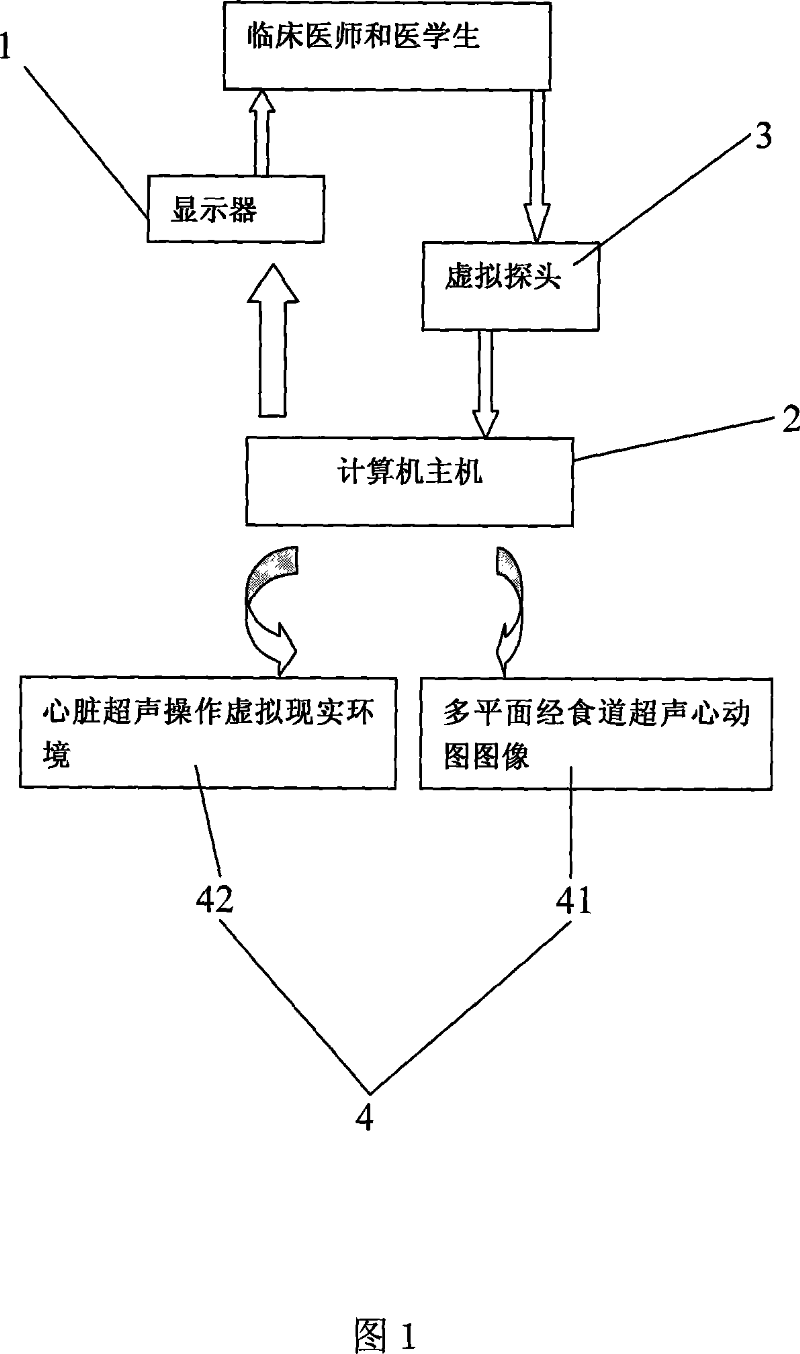
[0027]The establishment of a virtual transesophageal echocardiography system must meet the following conditions: First, it is necessary to establish a thin-section anatomical control corresponding to any orientation of the transesophageal echocardiographic view, so as to facilitate the physician's identification of the anatomical structure of the constantly changing ultrasonic view; Secondly, because transesophageal echocardiography can display the subtle anatomical structures in the heart, it is necessary to establish a high-quality three-dimensional visualization model of the heart through careful image segmentation technology; finally, the operation of transesophageal echocardiography is more complicated than that of transthoracic echocardiography. A virtual technology must be designed and studied to ensure that the virtual transesophageal echocardiography system has the characteristics of easy operation and realistic environment. At present, there are no reports on the rese...


PUM


Abstract

The invention discloses an ultrasonic cadiogram system and the realizing method through the esophagus virtually, wherein the system includes a display, a main frame connected to the display and a virtual probe connected to the main frame; a virtual multiple plane ultrasonic cadiogram system module via the esophagus is arranged in the main frame, wherein the system module includes a multiple plane ultrasonic cadiogram data base via the esophagus and a virtual reality environment for the heart ultrasonic operation; the system is applied in the clinic and the practical work, for solving the a plurality of problems which are not identified by the esophagus ultrasonic cadiogram image and are hard to popularize and master by the esophagus ultrasonic cadiogram, in addition, the clinicians and the medicos can grasp the anatomical structure of different esophagus depth and the angle via the ultrasonic cadiogram image on said system, which is favorable for the popularization of the ultrasonic cadiogram via the esophagus and advances the accuracy for diagnosing the heart disease via the esophagus ultrasonic cadiogram, thereby avoiding from adding the pain to the sufferer and reducing the incidence rate of the complication.

Description

technical field [0001] The invention relates to a medical imaging technology-ultrasound medical imaging, in particular to a virtual transesophageal echocardiography system and its implementation method. Background technique [0002] Ultrasound diagnosis of heart disease has always been the focus and difficulty in the field of ultrasound diagnosis. Transesophageal echocardiography (TEE) is a major advance in the field of cardiovascular disease diagnosis in recent years. It overcomes the shortcomings of traditional transthoracic echocardiography and provides a new means for the diagnosis of various heart and great vessel diseases. , especially the advent of multi-plane TEE probes, has made a qualitative leap in the development of TEE. Multiplanar TEE can play an important clinical role in the diagnosis, intraoperative monitoring, and postoperative follow-up of various heart diseases such as aortic dissection, congenital heart disease, coronary heart disease, and valvular hear...

Claims


Application Information

Patent Timeline
IPC IPC(8): A61B8/00A61B19/00G09B9/00G09B19/00A61B8/12
Inventor 郭燕丽张绍祥
Owner THE FIRST AFFILIATED HOSPITAL OF THIRD MILITARY MEDICAL UNIVERSITY OF PLA
Why Eureka
Try Eureka