Automatic imaging plane selection for echocardiography

an automatic selection and echocardiography technology, applied in the field of anatomy, can solve the problems of difficult to achieve high beam density and inaccurate quantification, and achieve the effects of high tissue contrast, fast diagnosis, and high resolution

Inactive Publication Date: 2015-01-08
KONINKLJIJKE PHILIPS NV
View PDF3 Cites 27 Cited by
  • Summary
  • Abstract
  • Description
  • Claims
  • Application Information

AI Technical Summary

Benefits of technology

[0005]The non-ionizing, high resolution and high tissue contrast advantages of echocardiography enable fast diagnosis and reliable guidance for interventional applications.

Problems solved by technology

This is difficult as the user needs to orient the ultrasound transducer probe to optimally capture the standard views (e.g. 2D images, X-planes which are two views that cross the apex (axially closest point) of the image, or selected volumetric acquisition).
In addition, the trade-off between beam density, volume size and frame rate in 3D echocardiography makes accurate quantification challenging when looking at complex structures such as the aortic root or aortic aneurysms.
Accurate measurements require high beam density which is hard to attain if imaging a large volume at high frame rate.

Method used


Image

  • Automatic imaging plane selection for echocardiography
  • Automatic imaging plane selection for echocardiography
  • Automatic imaging plane selection for echocardiography

Examples


Embodiment Construction

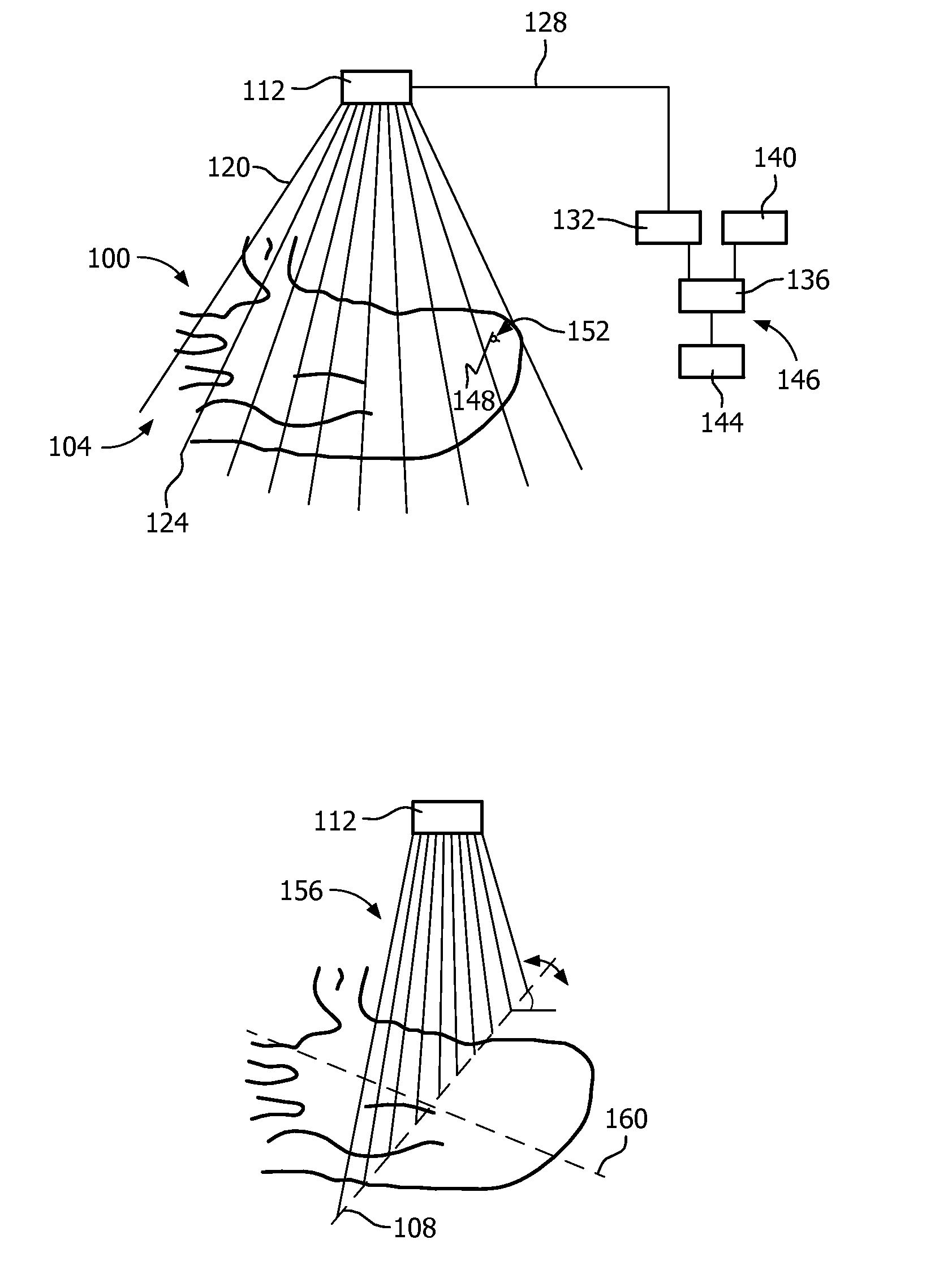
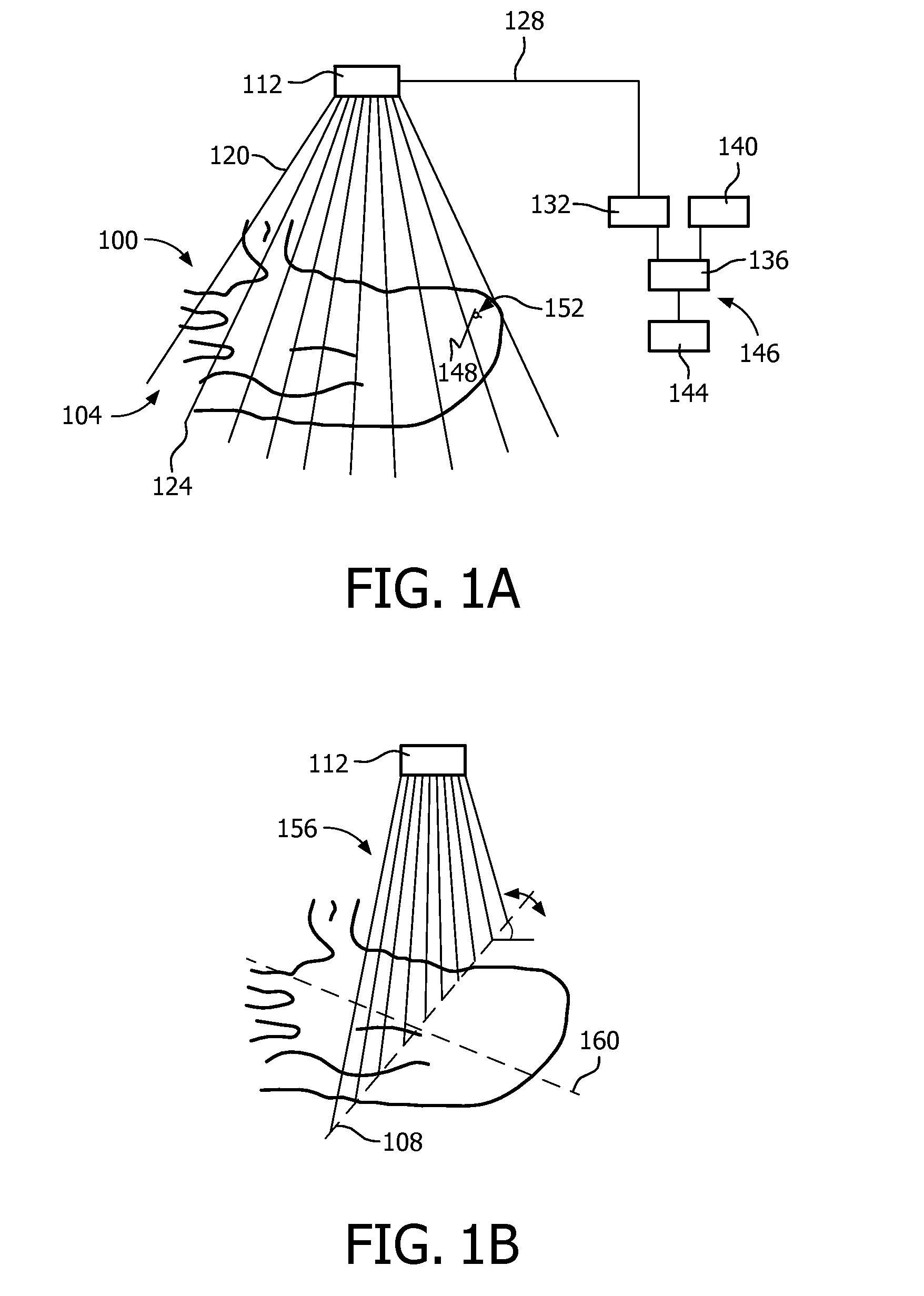
[0041]FIGS. 1A and 1B show, by way of illustrative and non-limitative example, live imaging of a volume 100 such as an entire heart 104 of a human or animal, that imaging being subsequently reduced to live imaging within a single imaging plane 108. The imaging, in both cases, is performed by a probe 112. For cardiac applications, the probe 112 can be a TEE probe, for intracorporeal use, or a TTE probe. The TEE probe is advanced down the esophagus into position for imaging. In the TTE case, an imaging end of the probe 112 will typically be handheld and controlled by a sonographer, cardiologist or radiologist. In the TEE case, the probe 112 will typically be controllable by one or more pull cables for steering and a multiplane probe will be rotatable within the esophagus manually or by a motor. This maneuvering can be done under image guidance afforded by an imaging window in the probe 112 through which ultrasound imaging is performed. Once the probe 112 is properly positioned, volume...


PUM


Abstract

Based on anatomy recognition from three-dimensional live imaging of a volume, one or more portions (204, 208) of the volume are selected in real time. In further real time response, live imaging or the portion(s) is performed with a beam density (156) higher than that used in the volume imaging. The one or more portion may be one or more imaging plane selected for optimal orientation in making an anatomical measurement (424) or display. The recognition can be based on an anatomical model, such as a cardiac mesh model. The model may be pre-encoded with information that can be associated with image locations to provide the basis for portion selection, and for placement of indicia (416, 420, 432, 436) displayable for initiating measurement within an image provided by the live portion imaging. A single TEE or TTE imaging probe (112) may be used throughout. On request, periodically or based on detected motion of the probe with respect to the anatomy, the whole process can be re-executed, starting back from volume acquisition (S508).

Description

FIELD OF THE INVENTION[0001]The present invention relates to imaging anatomy and, more particularly, to, based on the anatomy in the imaging, modifying the imaging.BACKGROUND OF THE INVENTION[0002]Accurate anatomical measurements are needed pre-treatment and for diagnosis of a number of cardiac conditions. The gold standard treatment for aortic stenosis is surgical replacement of the aortic valve. Open heart surgery is required, but is not an option for some elderly patients. A recently-developed enormously less invasive alternative is Transcatheter Aortic Valve Implantation (TAVI). Via a catheter, a replacement valve is advanced intravenously and disposed at the location of the current, faulty valve. In preparation, the diameter of the aortic valve annulus is determined. As another example, the diameter of the ascending aorta is calculated to assess the potential for an aneurysm.[0003]Medical imaging is a non-invasive method for making the anatomical measurements. In medical image ...

Claims


Application Information

Patent Timeline
Patent Type & Authority Applications(United States)
IPC IPC(8): A61B8/08A61B8/14
CPCA61B8/0883A61B8/145A61B8/5215A61B8/483A61B8/488A61B8/12A61B8/585G01S7/52085
Inventor RADULESCU, EMIL GEORGEWEESE, JUERGENSALGO, IVAN
Owner KONINKLJIJKE PHILIPS NV
Why Eureka
Try Eureka