Nanoparticle-based imaging agents for X-ray/computed tomography and methods for making same

a technology of computed tomography and nanoparticles, applied in the direction of pharmaceutical delivery mechanism, pigment treatment with organosilicon compounds, chemistry apparatus and processes, etc., can solve the problems of poor cost-effectiveness, difficult to target these agents to disease sites, and robust synthesis and instability

Active Publication Date: 2015-10-06
GE HEALTHCARE LTD
View PDF46 Cites 0 Cited by
  • Summary
  • Abstract
  • Description
  • Claims
  • Application Information

AI Technical Summary

Benefits of technology

[0016]The present invention uses a nanoparticle approach to deliver a relatively large number of high-density, highly-attenuating (radio-opaque molecular structures with effective atomic number greater than or equal to Z=34, the atomic number of selenium) atoms in elemental or molecular form to improve CT contrast enhancement. In some embodiments, the present invention provides for targeting of specific disease sites by the CT imaging agent. In some embodiments, the present invention provides for macrophage uptake of the CT imaging agent. In some embodiments, the present invention provides for a CT imaging agent with increased blood half-life.

Problems solved by technology

Additionally, such standard CT imaging agents are typically of low molecular weight, and they are known to clear from the human body very rapidly, making it difficult to target these agents to disease sites (Shi-Bao Yu and Alan D. Watson, Chem. Rev. 1999, 99, 2353-2377).
However, in such systems, only a relatively small number of heavy atoms may be delivered to / in the vicinity of the target tissues.
Although nanoparticles of elemental (zerovalent) metal species have the highest density (number of heavy metal atoms / volume), they suffer from issues such as robust synthesis and instability due to oxidation.
Nanoparticles of inert metals such as gold (e.g., such as described in WO 03 / 075961 A2) can overcome these issues, but are not very cost effective.

Method used


Image

  • Nanoparticle-based imaging agents for X-ray/computed tomography and methods for making same
  • Nanoparticle-based imaging agents for X-ray/computed tomography and methods for making same
  • Nanoparticle-based imaging agents for X-ray/computed tomography and methods for making same

Examples


example 1

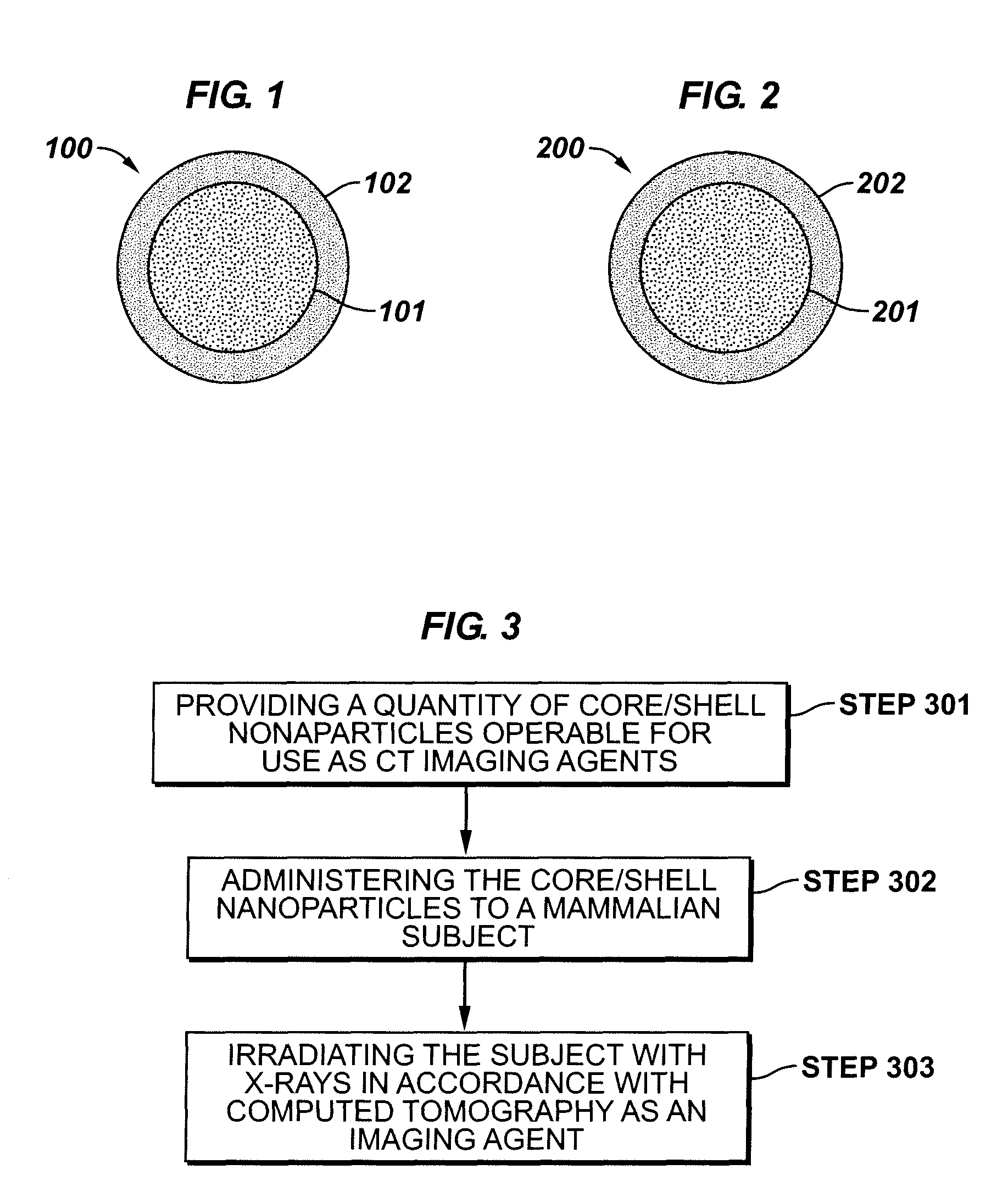
[0061]This Example serves to illustrate how a CT imaging agent can be prepared, in accordance with some embodiments of the present invention. In this particular Example, a passive shell is linked to an active core of hafnium oxide.

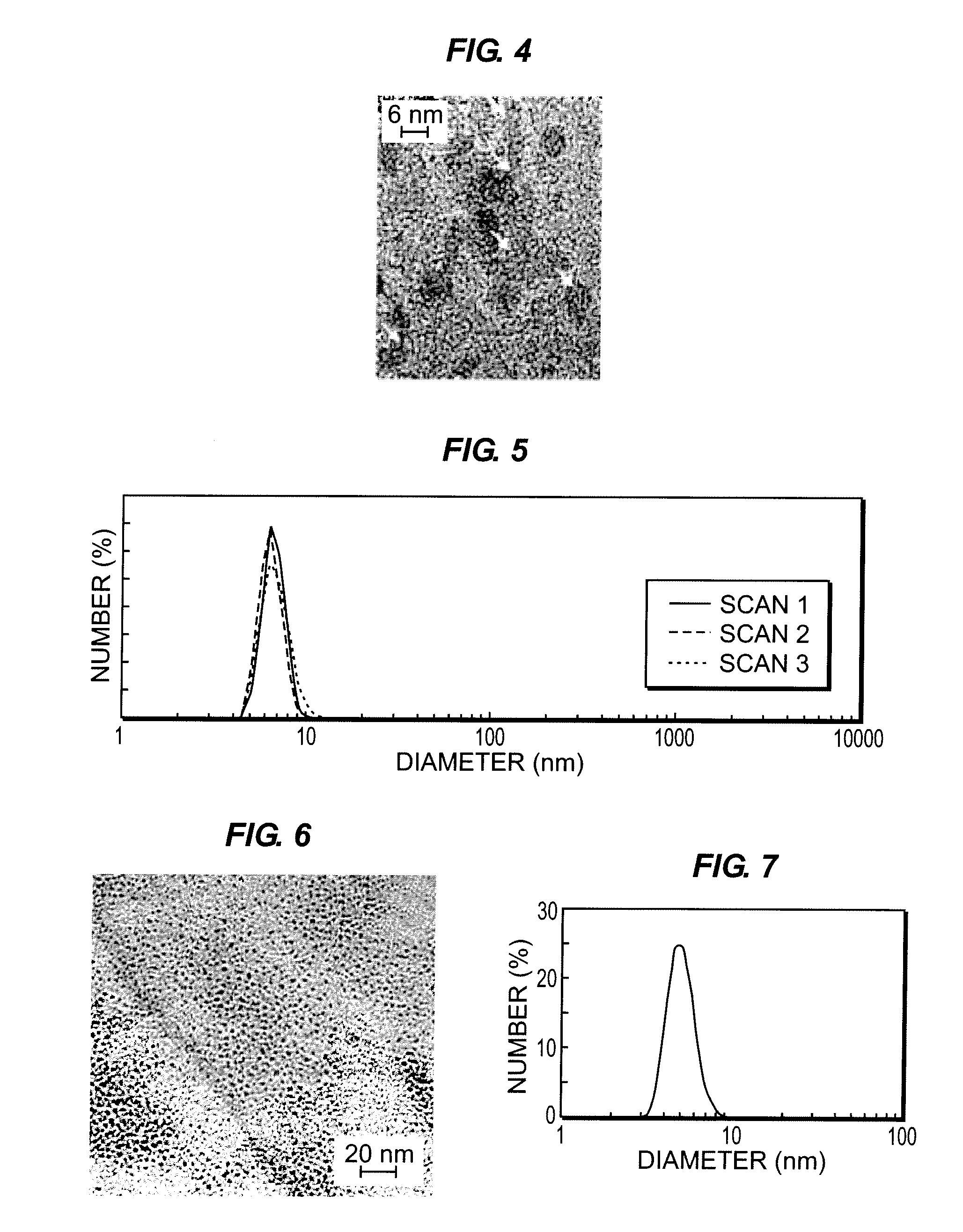
[0062]Hafnia nanocrystals (HfO2) were prepared from a suspension of hafnium oxychloride in ethanol. An organosilane-based coating was applied as follows: dilute 3-glycidoxypropyl(trimethoxysilane), GPTS was diluted using butanol (volume ratio 1:0.5) and pre-hydrolyzed step by addition of 0.1M HCl keeping the molar ratio of GPTS:H2O at 1:0.5. The resulting solution was subjected to vigorous stirring overnight at room temperature, then loaded with HfO2 nanocrystals. See Ribeiro et al., Appl. Phys. Lett. 2000, 77 (22), 3502-3504. FIG. 4 is a TEM image of active core / passive shell nanoparticles comprising a hafnium oxide core and polymeric shell, in accordance with some embodiments of the present invention.

example 2

[0063]This Example serves to illustrate how a CT imaging agent can be prepared, in accordance with some embodiments of the present invention. In this particular Example, a polymeric passive shell is linked to an active core of tantalum oxide. Further, in this particular Example, the shell formed about the core after the core is formed.

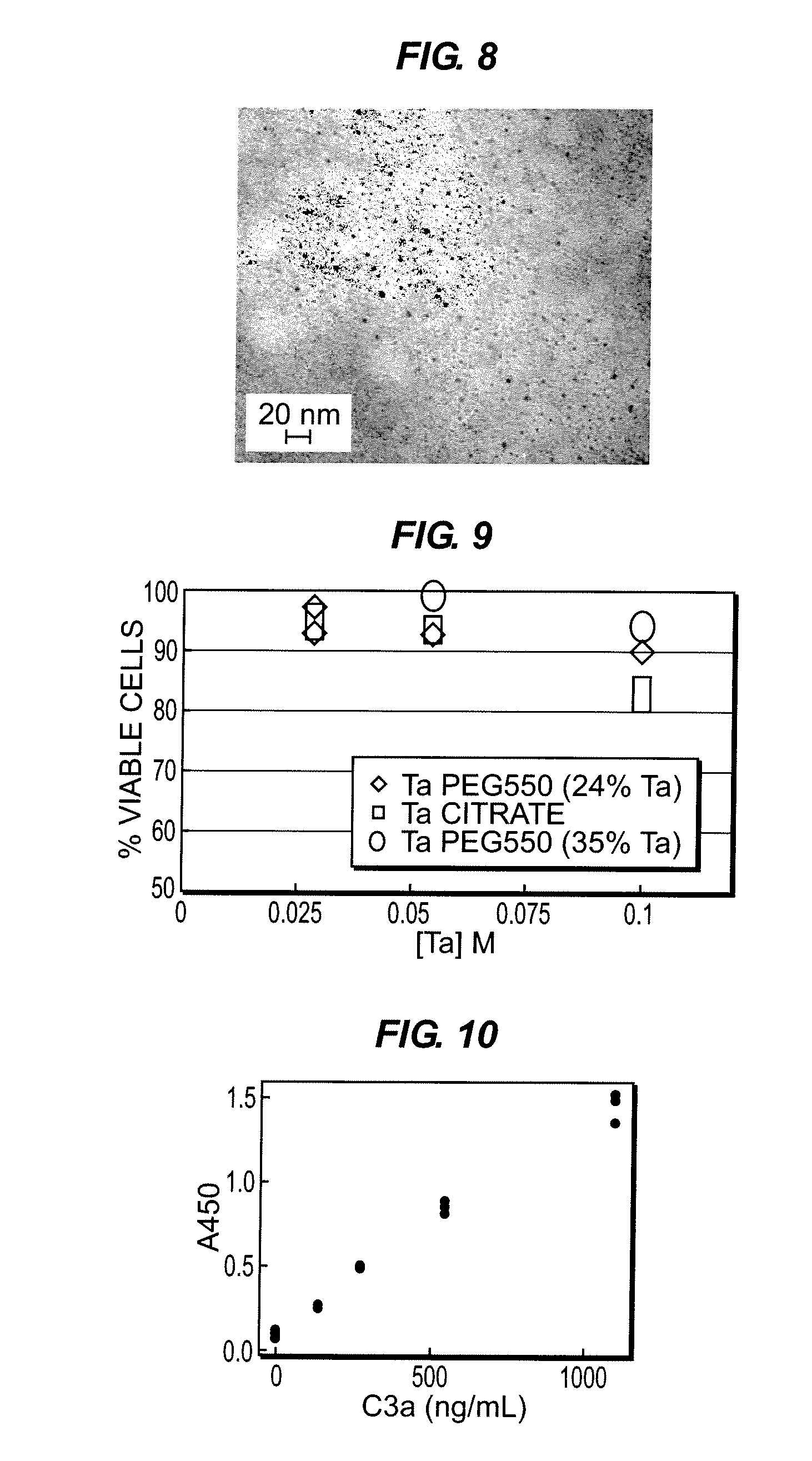
[0064]This example illustrates preparation of Ta2O5 nanoparticles for X-ray imaging: 34 ml of n-propanol, 0.44 ml isobutyric acid and 0.5 ml deuterium oxide were combined under nitrogen in the order specified and stirred for 30 minutes at room temperature. Tantalum ethoxide (1.87 g) was added in a drop-wise manner, albeit rapidly, and stirring continued under nitrogen for 18 hours. The tantalum ethoxide contains Ta(V), that is tantalum in the +5 valence state, a non-zero valence state. 2-[Methoxy(poly-ethylenoxy)propyl]trimethoxysilane (PEGsilane550, 4.832 g) was added to the stirred mixture as a 40 ml solution in n-propanol and the reaction was reflux...

example 3

[0066]This Example serves to illustrate how a CT imaging agent can be prepared, in accordance with some embodiments of the present invention. In this particular Example, a polymeric passive shell is linked to an active core of tantalum oxide. Further, in this particular Example, the shell formed about the core after the core is formed.

[0067]This example illustrates preparation of Ta2O5 nanoparticles for X-ray imaging: 34 ml of n-propanol, 0.44 ml isobutyric acid, and 0.5 ml deuterium oxide were combined under nitrogen in the order specified and stirred for 30 minutes at room temperature. Tantalum ethoxide (1.87 g) was added in a drop-wise manner, albeit rapidly, and stirring continued under nitrogen for 18 hours. The tantalum ethoxide contains Ta(V), that is tantalum in the +5 valence state, a non-zero valence state. Next, diethylphosphatoethyltriethoxysilane (PHS, 3 g) was added to the mixture as a 40 ml solution in n-propanol and the reaction was refluxed for 1.5 hours in air. Onc...


PUM

PropertyMeasurementUnit
mean diameteraaaaaaaaaa
energyaaaaaaaaaa

Abstract

The present invention is generally directed to core / shell nanoparticles, wherein such core / shell nanoparticles comprise a nanoparticle core and a nanoshell disposed about the nanoparticle core such that, in the aggregate, they form a core / shell nanoparticle that is operable for use as an imaging agent in X-ray / computed tomography (CT). Typically, such core / shell nanoparticle-based X-ray CT imaging agents further comprise a targeting species for targeting the imaging agent to diseased sites. Included herein are methods for forming such agents, comprising forming an ensemble of core / shell nanoparticles, wherein the mean diameter of the ensemble of core / shell nanoparticles is selected so as to render the nanoparticles in the ensemble substantially clearable by a mammalian kidney.

Description

RELATED APPLICATIONS[0001]This application is a continuation-in-part of U.S. patent application Ser. No. 11 / 627,529, filed Jan. 26, 2007, which itself is a continuation-in-part of U.S. patent application Ser. No. 11 / 265,728, filed Nov. 2, 2005, entitled “Nanoparticle-based Imaging Agents for X-Ray / Computer Tomography,” each of which is hereby incorporated herein by reference.TECHNICAL FIELD[0002]The present invention relates generally to imaging agents for use in X-ray / computed tomography, and more specifically to nanoparticle-based imaging agents and methods for making same.BACKGROUND INFORMATION[0003]Iodinated benzoic acid derivatives continue to serve as standard X-ray / computed tomography (CT) imaging agents, despite the risk factors and side effects associated with intravenous iodine injection. Additionally, such standard CT imaging agents are typically of low molecular weight, and they are known to clear from the human body very rapidly, making it difficult to target these agen...

Claims


Application Information

Patent Timeline
Patent Type & Authority Patents(United States)
IPC IPC(8): A61K49/04B82Y30/00C01G27/02C01G35/00C01G3/12B82Y5/00C09C3/12
CPCA61K49/0423B82Y5/00B82Y30/00C01G27/02C01G35/00C09C3/12C01P2004/04C01P2004/64
Inventor BONITATIBUS, JR., PETER JOHNDEMOULPIED, DAVID CHENEYTORRES, ANDREW SOLIZKULKARNI, AMIT MOHANCOLBORN, ROBERT EDGARBUCKLEY, PAUL WILLIAM
Owner GE HEALTHCARE LTD
Why Eureka
Try Eureka