Microvitreoretinal surgery blades

a micro-vitreoretinal and surgical technology, applied in the field of cutting tissue, can solve the problems of prolonged (months to years) and high cost of using these conventional treatments for retinal vein occlusion treatment, and achieve the effect of enhancing hemostasis or chorioretinal anastomosis

Inactive Publication Date: 2016-04-12
LUTTRULL JEFFREY K
View PDF10 Cites 0 Cited by
  • Summary
  • Abstract
  • Description
  • Claims
  • Application Information

AI Technical Summary

Problems solved by technology

Retinal vein occlusions are a common cause of visual loss.
Using these conventional treatments for retinal vein occlusion treatment is typically prolonged (months to years) and expensive.
Currently, there is no ideal tool for performing the critical part of the MTC procedure, which is the transvenous chorioretinotomies themselves.
Most modern vitreous surgery is performed with 23 or 25 gauge trocar systems, and the prior art MVR blades typically available for such procedures do not function well in MTCs.
The drawbacks of such prior art MVR blades include: (a) the blade tips are too narrow, making it difficult to center the blade on the target vein to achieve complete transection; (b) the side edges of the blades are too dull, causing them to displace rather than transect the target vein; and (c) the blade tips are too long, resulting in increased risk of scleral perforation and globe perforation, particularly in myopic patients with thin sclera.
However, the MTC incision it creates is unnecessarily large and use of the 20 gauge blade requires creation of an additional incision in the eye, which adds steps to the procedure, prolonging the surgery and increases the risk of complications.

Method used


Image

  • Microvitreoretinal surgery blades
  • Microvitreoretinal surgery blades
  • Microvitreoretinal surgery blades

Examples


first embodiment

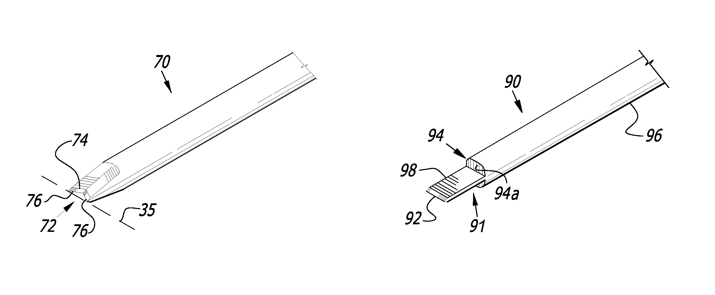
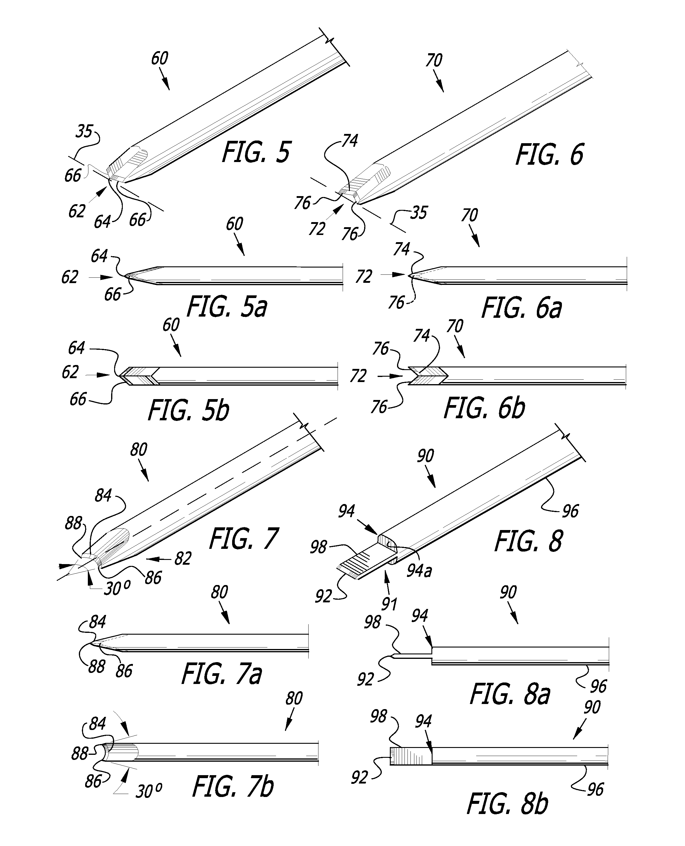
[0055]FIGS. 8, 8a and 8b depict a seventh preferred embodiment of the inventive MVR blade 90, sometimes referred to as a guarded blade or working tip 91. In this embodiment, the MVR blade 90 may have an edge similar to any of the earlier embodiments. FIG. 8 illustrates a thin chisel shape, i.e., a flat or horizontal, chisel-type edge 92, configured similarly to the first embodiment MVR blade 30. The distinguishing feature of this embodiment is the step-down 94, i.e., sudden reduction in cross-sectional width, from the shaft 96 to the blade body 98. The step-down 94 operates to limit the depth of penetration to reduce the risk of scleral perforation or other negative effects of over-insertion. The step-down 94 provides a lip or stop ledge 94a on the end of the shaft 96 that contacts but does not cut or penetrate tissue. It is this contact that stops the cutting motion and prevents over-insertion. Although the blade body 98 may be longer, it is preferably approximately 1 mm-2 mm in le...

sixth embodiment

[0056]FIG. 9 illustrates an environmental view of an MVR blade 80 of the present invention, in this instance the sixth embodiment, penetrating an eye 100 through an incision 102. This and any other similar procedure may be performed with a blade of any other embodiment described herein. In accessing the retina of an eye for performing a transvenous chorioretinotomy or other similar procedures, an MVR blade must pass through the eye 100 in this way and access the retina at the back of the eye 100.

[0057]FIGS. 10 through 17 illustrate various views of an MVR blade 80 performing such a transvenous chorioretinotomy procedure. In these illustrations, the interior 100a of the eye is on top and the exterior 100b of the eye is on bottom.

[0058]In FIGS. 10 and 11, the blade 80 is approaching the back of the eye 100, which is comprised of various layers, including the retina 104, the choroid 106 and the sclera 108. The retina 104 contains a vast number of veins 110, of which only one is shown i...


PUM


Abstract

A micro-vitreoretinal (MVR) blade for incising tissue for a transvenous chorioretinotomy and similar procedures includes a shaft with a working tip having a chisel-type edge on its end or leading edge. Various embodiments of the inventive MVR blade have different chisel-type edge structures, such as a latitudinal chisel, an angled chisel, a chevron, a reverse chevron, a concave semi-lunar and a guarded or step-down working tip. The MVR blade may also include drug / chemical coatings, electrification or freezing to promote hemostasis or chorioretinal anastomosis formation.

Description

BACKGROUND OF THE INVENTION[0001]The present invention relates generally to incising tissue and in particular to micro-vitreoretinal surgery blades. In just one example, micro-vitreoretinal (MVR) blades are used to incise tissue for a transvenous chorioretinotomy as treatment for retinal vein occlusion and other indications. Retinal vein occlusions are a common cause of visual loss. Currently, the primary treatments are macular laser (for branch retinal vein occlusion) and pharmacologic agents (for branch and central retinal vein occlusion). These treatments address the side-effects of the retinal vein occlusion, including macular edema and ocular neovascularization. Using these conventional treatments for retinal vein occlusion treatment is typically prolonged (months to years) and expensive.[0002]Recently, a new procedure for treating retinal vein occlusions was reported that involves bypassing the occlusion through surgery to create a chorioretinal anastomosis. Unlike previous at...

Claims


Application Information

Patent Timeline
Patent Type & Authority Patents(United States)
IPC IPC(8): A61B17/32A61B17/34A61F9/007
CPCA61B17/3417A61F9/00736A61B2017/3454
Inventor LUTTRULL, JEFFREY K.
Owner LUTTRULL JEFFREY K
Why Eureka
Try Eureka