Real-time lesion formation assessment

a real-time assessment and lesions technology, applied in the field of real-time assessment of lesions, can solve the problems of insufficient creation of reversible lesions to achieve electrical isolation, insufficient cryoballoon temperature, and high cost of operation

Active Publication Date: 2016-10-18
MEDTRONIC CRYOCATH LP
View PDF23 Cites 33 Cited by
  • Summary
  • Abstract
  • Description
  • Claims
  • Application Information

AI Technical Summary

Problems solved by technology

The creation of reversible lesions may not be sufficient to achieve electrical isolation and, as a result, atrial fibrillation may be likely to reoccur.
Additionally, even if the PV is completely occluded, suboptimal operation of the cryoablation system may result in cryoballoon temperatures that are not low enough, or not applied for a sufficient amount of time, to create permanent lesions in the target tissue.
However, each of these methods has its drawbacks.
This post-procedural medical imaging does not provide real-time lesion assessment and / or prevention of injury to non-target tissue.
Further, measuring the temperature of target tissue during a procedure may be difficult or impossible, and methods that measure temperature within the cryoballoon to approximate the temperature of treated tissue may not take into account the tissue type and response to treatment, and can be very inaccurate.
Likewise, temperature-time assessment methods may be based on a one-size-fits all model (for example, an Arrhenius-like equation) that does not take into account the type and depth of tissue, and may be subject to noise in the temperature data.
Further, the cell-death models may not be very robust, may over- or under-estimate the effects an ablation procedure has on the target tissue, and different models may have to be used for different tissue.
None of the methods described above provides real-time temperature and occlusion feedback that can be used to assess lesion formation in real time.

Method used


Image

  • Real-time lesion formation assessment
  • Real-time lesion formation assessment
  • Real-time lesion formation assessment

Examples


first embodiment

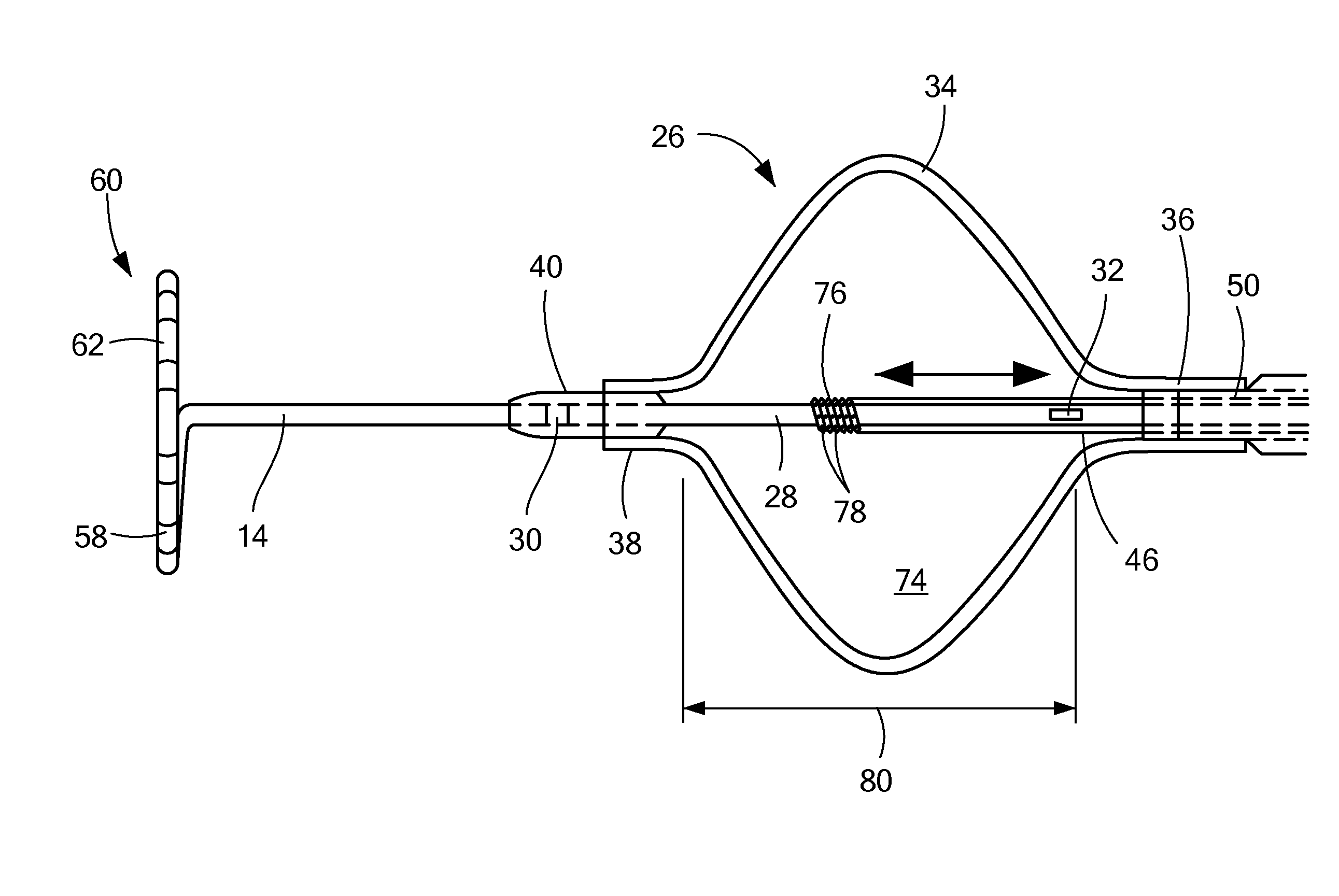
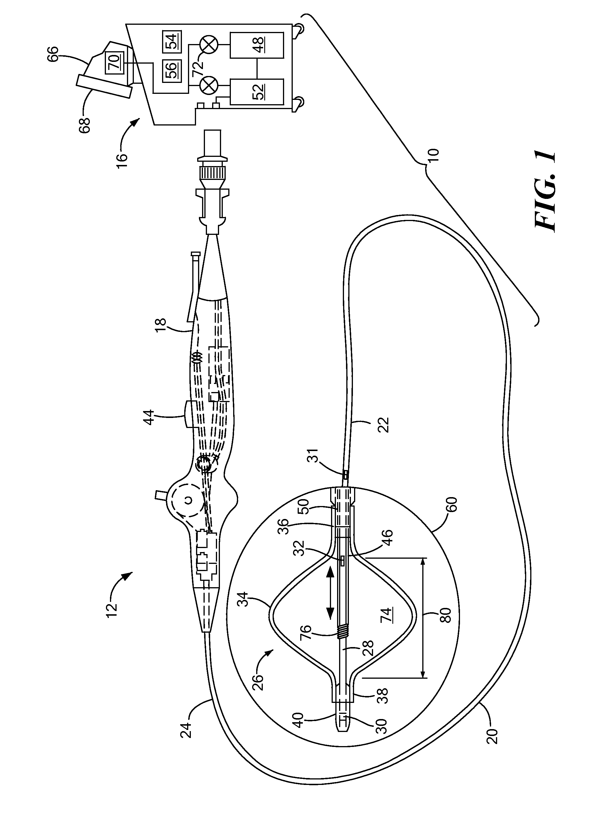
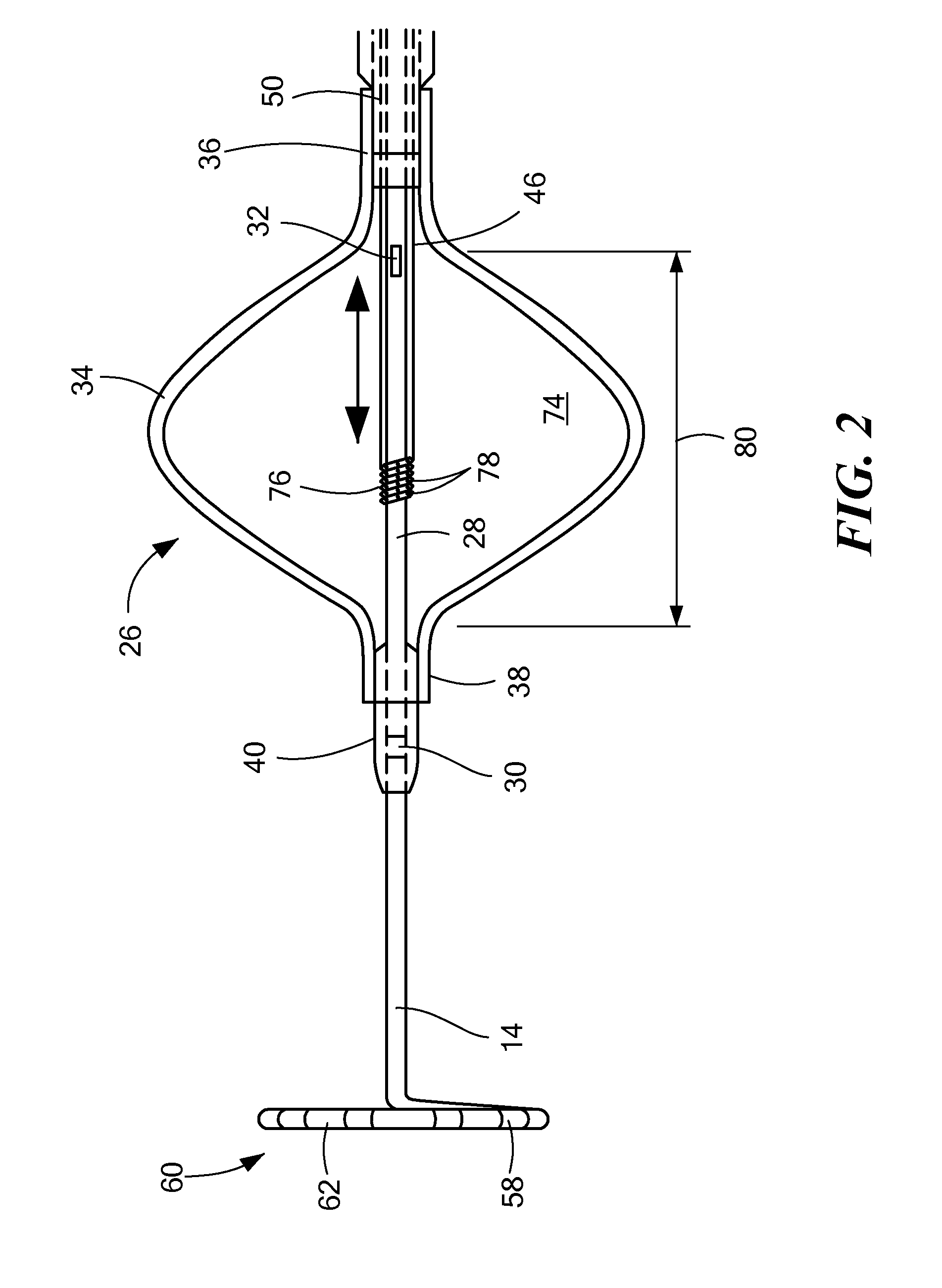
[0036]Referring now to FIGS. 3A-3E, different embodiments of ways in which a temperature sensor may be coupled to a cryotreatment catheter. In the first embodiment shown in FIG. 3A, the distal temperature sensor 30 may be coupled to an outer surface of the nose 82. Although FIG. 3A shows the distal temperature sensor 30 protruding from the outer surface 84 of the nose 82, the figures may not be drawn to scale and the temperature sensor 30 may be thin enough that the sensor 30 lies flush or substantially flush with the outer surface 84 of the nose 82. Alternatively, the sensor 30 may be placed within a recessed area of the nose 82, thereby causing the sensor 30 to lie flush or substantially flush with the outer surface 84 of the nose 82. The sensor 30 may be a discrete sensor (that is, not be configured as a band or array of sensors) and may include one or more wires 86 that transmit signals (for example, temperature measurements by the sensor 30) to the console, and the wires 86 may...

second embodiment

[0037]In the second embodiment shown in FIG. 3B, the distal sensor 30 may be a discrete sensor and may be disposed within the guide wire lumen 28, but may protrude from the outer surface 84 of the nose 82. Alternatively, the temperature sensor 30 may be thin enough that the sensor 30 lies flush or substantially flush with the outer surface 84 of the nose 82 (as shown in FIG. 3B). The one or more wires 86 may be disposed within the guide wire lumen 28, and may travel the length of the device within the guide wire lumen 28 to the handle 18, and may also be disposed within the handle 18 and in electrical communication with the console as described in FIG. 3A.

third embodiment

[0038]In the third embodiment shown in FIG. 3C, the distal sensor 30 may be entirely disposed within the guide wire lumen 28, such that temperature measurements may be taken through the material of the guide wire lumen. In such an embodiment, the thickness of the wall of the guide wire lumen 28 may be thinnest proximate the distal sensor 30 to facilitate temperature measurement of the outside environment. The sensor's electrical wires 86 may be disposed within the guide wire lumen 28, and may travel the length of the device within the guide wire lumen 28 to the handle 18, and may also be disposed within the handle 18 and in electrical communication with the console as described in FIG. 3A.


PUM


Abstract

A method and system for creating permanent lesions in an area of target tissue, such as tissue at or proximate a junction between a pulmonary vein and the left atrium. The method may generally include positioning a medical device in contact with a pulmonary vein ostium, ablating the tissue, and recording a plurality of temperature measurements from one or more of three temperature sensors. The device may include an occlusion element in communication with a coolant source, a first sensor located distal of the occlusion element, a second sensor located proximal of the occlusion element, and a third sensor located in the occlusion element. One or more temperature measurements may be compared with each other to assess occlusion of the pulmonary vein, and / or may be compared with a set of reference temperatures to predict a real-time temperature within the target tissue.

Description

CROSS-REFERENCE TO RELATED APPLICATION[0001]n / aSTATEMENT REGARDING FEDERALLY SPONSORED RESEARCH OR DEVELOPMENT[0002]n / aFIELD OF THE INVENTION[0003]The present invention relates to a method and system for the real-time assessment of lesions formed during a cryoablation procedure.BACKGROUND OF THE INVENTION[0004]A cardiac arrhythmia is a condition in which the heart's normal rhythm is disrupted. Certain types of cardiac arrhythmias, including ventricular tachycardia and atrial fibrillation, may be treated by ablation (for example, radiofrequency (RF) ablation, cryoablation, ultrasound ablation, laser ablation, microwave ablation, and the like), either endocardially or epicardially.[0005]Procedures such as pulmonary vein isolation (PVI) and pulmonary vein antrum isolation (PVAI) are commonly used to treat atrial fibrillation. These procedures generally involve the use of a cryogenic device, such as a catheter, which is positioned at the ostium of a pulmonary vein (PV) such that any blo...

Claims


Application Information

Patent Timeline
Patent Type & Authority Patents(United States)
IPC IPC(8): A61B18/02A61B18/00
CPCA61B18/02A61B2018/0022A61B2018/00375A61B2018/00577A61B2018/00791A61B2018/00797A61B2018/00839A61B2018/0212A61B2018/0293
Inventor WITTENBERGER, DANLALONDE, JEAN-PIERREHOWARD, STEPHEN A.
Owner MEDTRONIC CRYOCATH LP
Why Eureka
Try Eureka