Wound healing metakaryotic stem cells and methods of use thereof

a technology of metakaryotic stem cells and wound healing, applied in biochemistry apparatus and processes, instruments, material analysis, etc., can solve the problems of affecting the quality of life, affecting the and the improvement of these interventions may be only temporary, and achieve the effect of rapid appearance of metakaryotic cells

Active Publication Date: 2016-11-22
MASSACHUSETTS INST OF TECH
View PDF35 Cites 0 Cited by
  • Summary
  • Abstract
  • Description
  • Claims
  • Application Information

AI Technical Summary

Benefits of technology

[0006]In one aspect, the invention provides methods of identifying the stem cells of wound healing generally by recognition of cells and / or syncytia with bell-shaped nuclei actively dividing by both symmetric and asymmetric amitoses. Examples include the rapid appearance of metakaryotic cells at wound edges after corneal transplants and the appearance of metakaryotic tubular syncytia during healing of skeletal muscle wounds.

Problems solved by technology

Wounds and wound healing disorders are pervasive and detract from the quality of life.
Wounds that sever nerve communication result in paralysis.
The improvements from these interventions may be only temporary, however, since wound healing disorders are commonplace and can give rise to complications such as restenosis, a re-narrowing of vessels following surgical intervention caused by a rapid increase in the number of smooth muscle cells of the vessel wall.
Unfortunately, it has been demonstrated that such antigenic markers or stem cells are widely distributed on and among cells that are not stem cells.
Said markers are useful in enriching stem cells from cell mixtures but do not permit isolation and observation of stem cells specifically.
These studies, however, do not demonstrate a role for the cells with stem-like properties in any disease.
In particular, Pasquinelli et al., did not observe symmetric or asymmetric amitoses of metakaryotes in vascular walls and did not observe creation of smooth muscle cells by asymmetric amitoses of metakaryotic cells.

Method used


Image

  • Wound healing metakaryotic stem cells and methods of use thereof
  • Wound healing metakaryotic stem cells and methods of use thereof
  • Wound healing metakaryotic stem cells and methods of use thereof

Examples


example 1

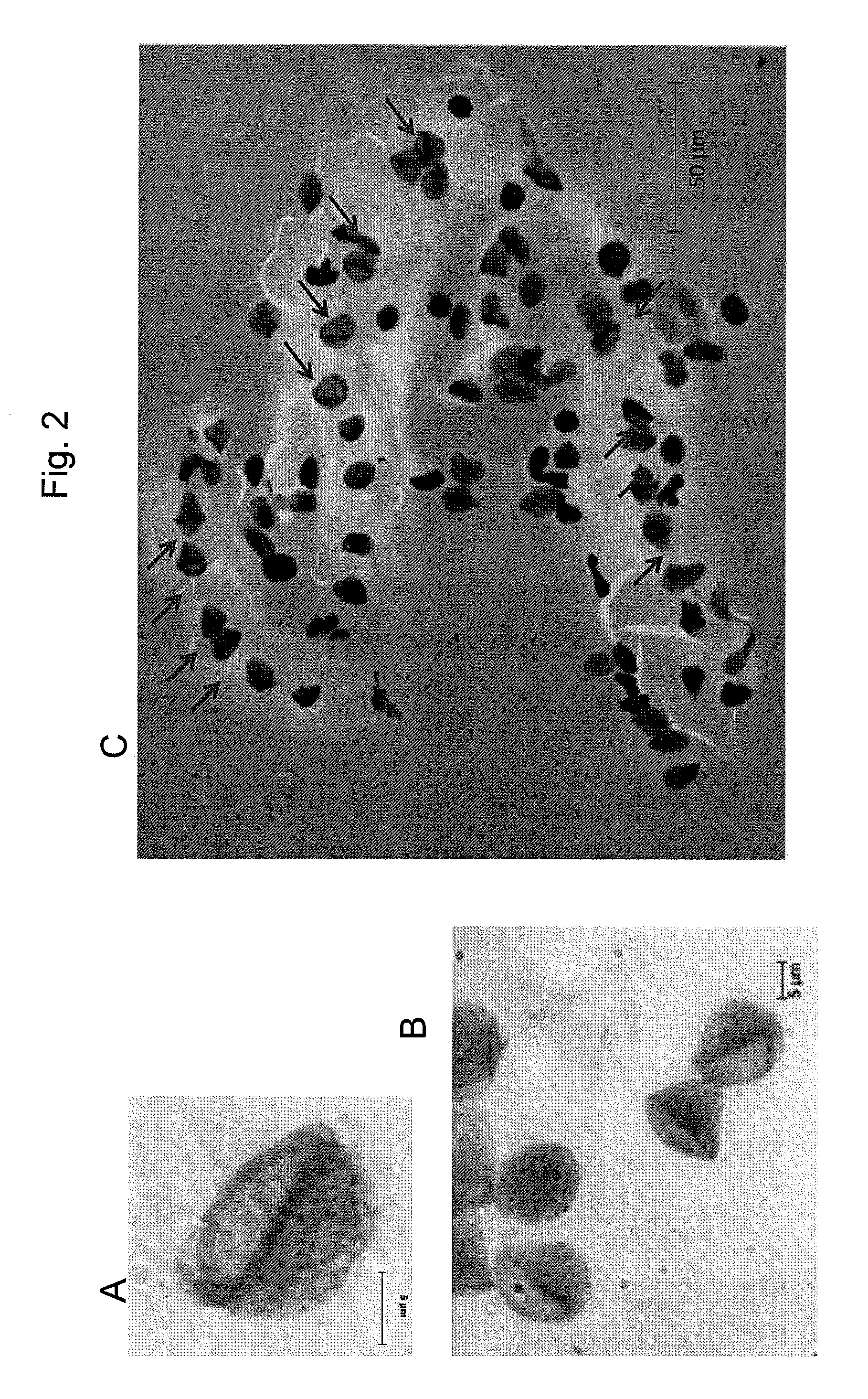
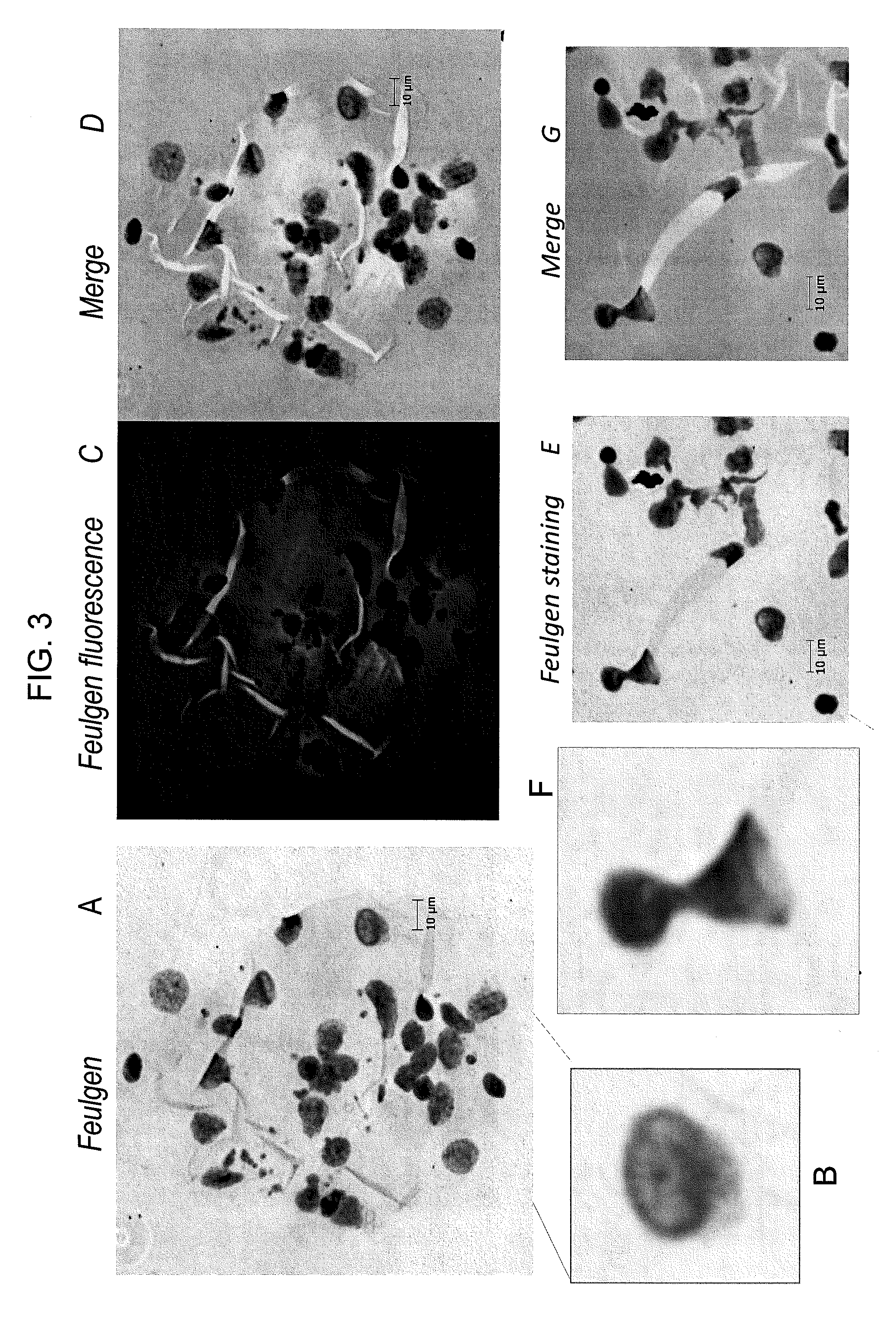
Visualization of Metakaryotic Cells in Fetal Kidney Artery

[0113]The tissues shown in FIGS. 2-4 were prepared for visualization of the metakaryotic stem cells substantially as described above. The tissues were obtained from human fetal kidney artery.

example 2

Visualization of Metakaryotic Cells in a Patient with Post-Transplant Restenosis

[0114]A child 2 years old who was recently transplanted, suffered rejection, and over 1 month had rapid progression of diffuse coronary atherosclerosis. The child suffered a cardiac arrest because of this rapidly progressing coronary disease. The child was stabilized on emergent cardiopulmonary bypass support for 6 days until a heart became available. The child's diseased heart was explanted and a new one was provided. FIG. 6 and FIG. 7 include micrographs of freshly-fixed tissues prepared from this subject. FIGS. 6C-D are micrographs showing major blood vessels in pig.

example 3

Vasculogenesis and Bladder Polyp Catheter Injury

[0115]FIGS. 2-4 illustrate vasculogensis. FIG. 17B includes a micrograph of a bladder polyp injury.


PUM

PropertyMeasurementUnit
diametersaaaaaaaaaa
diametersaaaaaaaaaa
thicknessaaaaaaaaaa

Abstract

The invention provides methods of identifying wound healing metakaryotic stem cells, identifying molecules to modulate proliferation and / or migration of metakaryotic stem cells and molecules to treat wound healing disorders, such as blood vessel wound healing disorders, including restenosis. The invention also provides methods of diagnosing and treating wound healing disorders, such as blood vessel wound healing disorders and also of treating wounds by the use of metakaryotic stem cell transplant(s).

Description

RELATED APPLICATION(S)[0001]This application is the U.S. National Stage of International Application No. PCT / US2011 / 057513, filed Oct. 24 2011, which designates the U.S., published in English, and claims the benefit of U.S. Provisional Application No. 61 / 406,468, filed on Oct. 25, 2010. The entire teachings of the above applications are incorporated herein by reference.BACKGROUND OF THE INVENTION[0002]Wounds and wound healing disorders are pervasive and detract from the quality of life. Wounds that sever nerve communication result in paralysis. Wound healing disorders afflict wounds in organs and tissues after traumatic injuries and infection and equally include wounds and wound-like conditions following surgical interventions as is frequently required for vascular diseases arising from progressive disorders such as atherosclerosis. For example, vascular diseases, such as coronary or carotid artery disease, affect millions of individuals in the U.S. and many more worldwide. See, e.g...

Claims


Application Information

Patent Timeline
Patent Type & Authority Patents(United States)
IPC IPC(8): C12Q1/02G01N33/68G01N33/50
CPCC12Q1/025G01N33/5058G01N33/6893G01N33/50G01N33/68
Inventor GOSTJEVA, ELENA V.THILLY, WILLIAM G.
Owner MASSACHUSETTS INST OF TECH
Why Eureka
Try Eureka