Spatial biomarker of disease and detection of spatial organization of cellular receptors

a cellular receptor and disease biomarker technology, applied in the field of live cell assays, can solve the problems of deconstructing the function of cell-surface signaling molecules, unable to elucidate in vivo receptor function and behavior, and more complicated conditions of diseased tissu

Active Publication Date: 2017-07-18
RGT UNIV OF CALIFORNIA
View PDF18 Cites 1 Cited by
  • Summary
  • Abstract
  • Description
  • Claims
  • Application Information

AI Technical Summary

Problems solved by technology

A more complicated condition is diseased tissue.
Deconstructing what cell-surface signaling molecules do after binding an ligand is a challenge because of the chemical heterogeneity of the cell membrane and the complexity of effecting cell-cell contact in an in vitro system.
While genetic screening and protein expression profiling can yield information, they fall short of elucidating in vivo receptor function and behavior.
These current techniques are usually indirect by design with respect to activation on the cell surface, and cannot be expected to perform beyond their limits, especially with regard to the challenge of decoding the complex events that activate the cell from a receptor at the surface.

Method used


Image

  • Spatial biomarker of disease and detection of spatial organization of cellular receptors
  • Spatial biomarker of disease and detection of spatial organization of cellular receptors
  • Spatial biomarker of disease and detection of spatial organization of cellular receptors

Examples


example 1

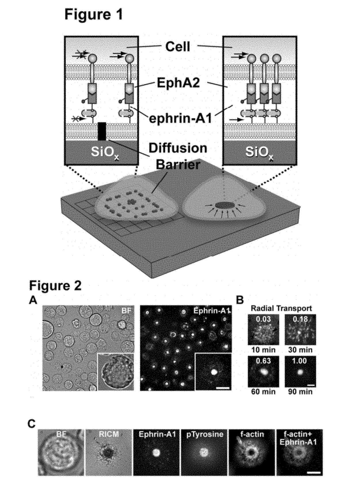
[0121]Many aspects of cancer result from aberrant signal transduction at the cell surface. Metastasis is one of the most deadly processes of cancer, and each of its phases (detachment, migration, invasion, growth, and survival) is regulated by cell-cell contact interactions and the associated signaling systems. For example, recent studies have found the EphA2 receptor tyrosine kinase (RTK) to be frequently over expressed and functionally altered in aggressive tumor cells (40% of breast cancers [B. L. Jackson, J. T. Groves, J. Am. Chem. Soc. 126, 13878 (2004)]), and that these changes promote metastatic character (FIG. 2A) [M. M. Davis et al., Annu. Rev. Biochem. 72, 717 (2003)]. EphA2 is one of the Eph receptors, which constitute the largest family of RTKs in the human genome and, together with their membrane-bound ephrin ligands, regulate a broad range of signaling processes at intercellular junctions. In addition to metastasis, Eph receptors are involved in oncogenic transformatio...

example 2

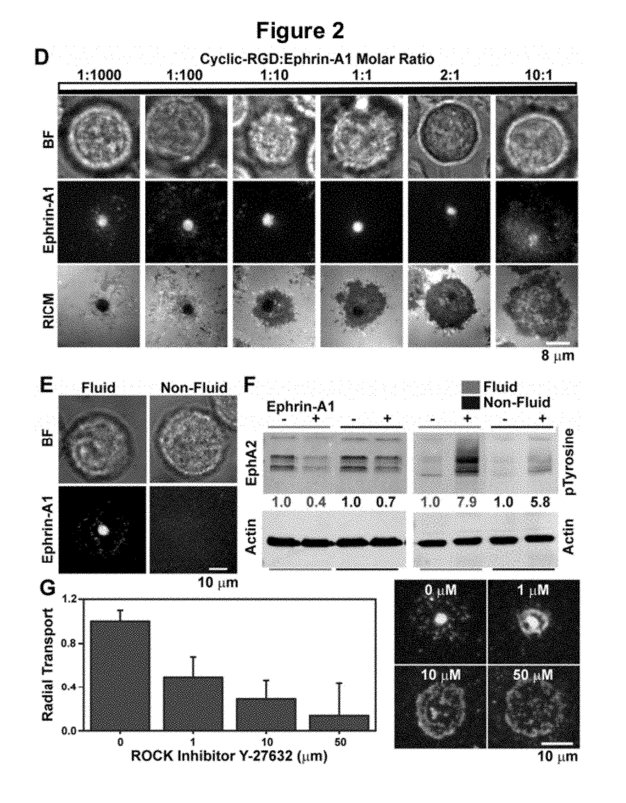
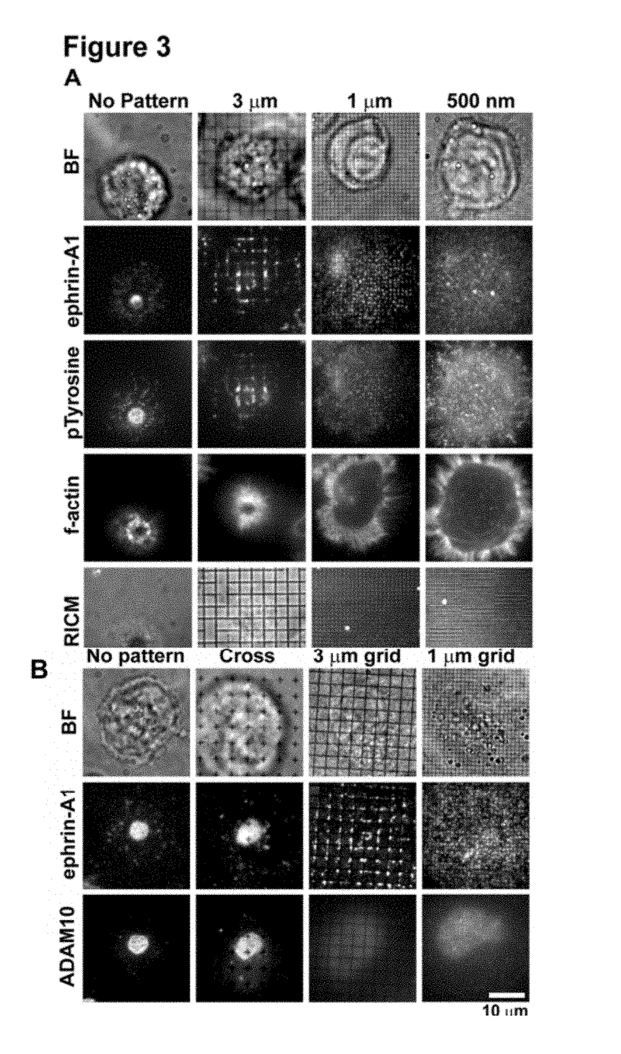
[0153]Activation of the EphA2 receptor tyrosine kinase by ephrin-A1 ligands presented on apposed cell surfaces plays important roles in development and exhibits poorly understood functional alterations in cancer. Here, we reconstitute this intermembrane signaling geometry between live EphA2-expressing human breast cancer cells and supported membranes displaying laterally mobile ephrin-A1. Receptor-ligand binding, clustering, and subsequent lateral transport within this junction are observed. EphA2 transport can be blocked by physical barriers nanofabricated onto the underlying substrate. This physical reorganization of EphA2 alters the cellular response to ephrin-A1, as observed by changes in cytoskeleton morphology and recruitment of the protease ADAM10. Quantitative analysis of receptor-ligand spatial organization across a library of 26 mammary epithelial cell lines reveals characteristic differences that strongly correlate with invasion potential. These observations reveal a mech...

example 3

[0158]The fluidity of the membrane is critical for observing CAD formation, and fully saturated 1,2-dipalmitoyl-sn-glycero-3-phosphocholine (DPPC) lipids that are non-fluid at 37° C. do not allow the coalescence of microclusters and their subsequent transport into the CAD (FIG. 2E).

[0159]To evaluate how the supported membrane affects EphA2 activation, we measured the degradation of EphA2, which is a direct consequence of ligand-induced receptor stimulation. Western blots measuring EphA2 degradation (FIG. 2D), indicate that membrane-bound ephrin-A1 activates EphA2 to the same degree as soluble ligand stimulation (1 ng / ml). However, ephrin-A1 anchored to non-fluid DPPC membranes can only activate EphA2 to ˜50% of the maximum achieved with fluid membranes (FIG. 2D). These results clearly indicate that the activity of chemically identical ligands can be modulated by altering membrane fluidity, thus suggesting an additional level of finely tuned responses in Eph-ephrin signaling.

[0160]Th...


PUM

PropertyMeasurementUnit
massaaaaaaaaaa
heightaaaaaaaaaa
heightaaaaaaaaaa

Abstract

A signature of a condition of a live cell is established in an assay that allows distribution of the receptors on the cell surface in response to binding a ligand. The receptors can be optically detected and quantified to provide a value for the condition, Test drugs can be screened for therapeutic potential in the assay: a potentially efficacious drug is identified by an ability to modulate an established signature. The receptor distribution signature can be corroborated with an mRNA expression profile of several genes, indicating, for example, metastasis.

Description

CROSS-REFERENCE TO RELATED APPLICATIONS[0001]The application claims priority to PCT International Application No. PCT / US2010 / 022671, filed Jan. 29, 2010, which claims priority to U.S. Provisional Patent Application No. 61 / 148,203, filed Jan. 29, 2009, and U.S. Provisional Patent Application No. 61 / 176,858, filed on May 8, 2009, each of which is hereby incorporated by reference in its entirety.[0002]The application hereby incorporates by reference in its entirety, co-pending U.S. patent application Ser. No. 12 / 161,553, filed on Jul. 21, 2008, entitled, “A Fluid Membrane-Based Ligand Display System for Live Cell Assays and Disease Diagnosis Applications.”STATEMENT OF GOVERNMENTAL SUPPORT[0003]This work was supported by Contract No. DE-AC02-05CH11231 awarded by the Department of Energy, by U.S. Department of Defense Breast Cancer Research Program Concept Award BC076701 under U.S. Army Medical Research Acquisition Activity No. W81XWH-08-1-0677, and by grants P50 CA 58207, and by the U54...

Claims


Application Information

Patent Timeline
Patent Type & Authority Patents(United States)
IPC IPC(8): G01N33/53C12Q1/68
CPCC12Q1/6886C12Q2600/136C12Q2600/156
Inventor SALAITA, KHALID S.NAIR, PRADEEP M.DAS, DEBOPRIYAGRAY, JOE W.GROVES, JOHN T.
Owner RGT UNIV OF CALIFORNIA
Why Eureka
Try Eureka