
Apparatus, systems, methods and computer-accessible medium for spectral analysis of optical coherence tomography images

a technology spectral analysis, which is applied in the field of spectral analysis of optical coherence tomography images, can solve the problems of difficult to definitively identify, difficult interpretation of images in other organ systems, and difficulty in identifying oct techniques, so as to improve the contrast of intracoronary oct images, reduce scattering effect, and increase accuracy and time

Active Publication Date: 2017-10-24
THE GENERAL HOSPITAL CORP
View PDF758 Cites 8 Cited by
  • Summary
  • Abstract
  • Description
  • Claims
  • Application Information

AI Technical Summary

Benefits of technology

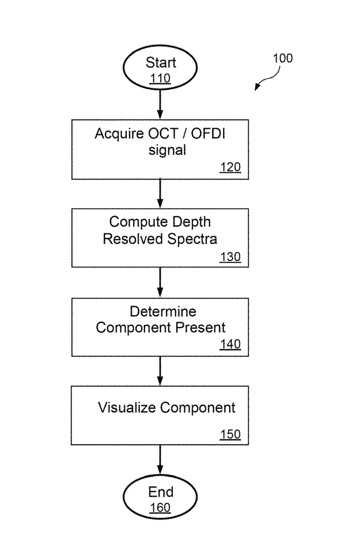
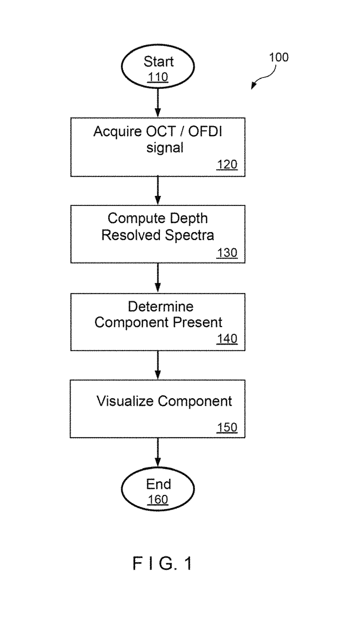
[0009]Herein, exemplary embodiments of systems, apparatus, methods and computer-accessible medium are described for depth resolved spectral analysis of OCT interferometric data and visualization methods for the identification of lipid rich regions. According to one exemplary embodiment, an analysis of inteferometric signals acquired (or previously acquired) with current coronary OCT catheters and systems can be utilized for spatial identification of lipid rich regions. The combined exemplary architectural and chemical analysis can improve contrast of intracoronary OCT images, which can increase the accuracy and time to which a user identifies diseased areas.
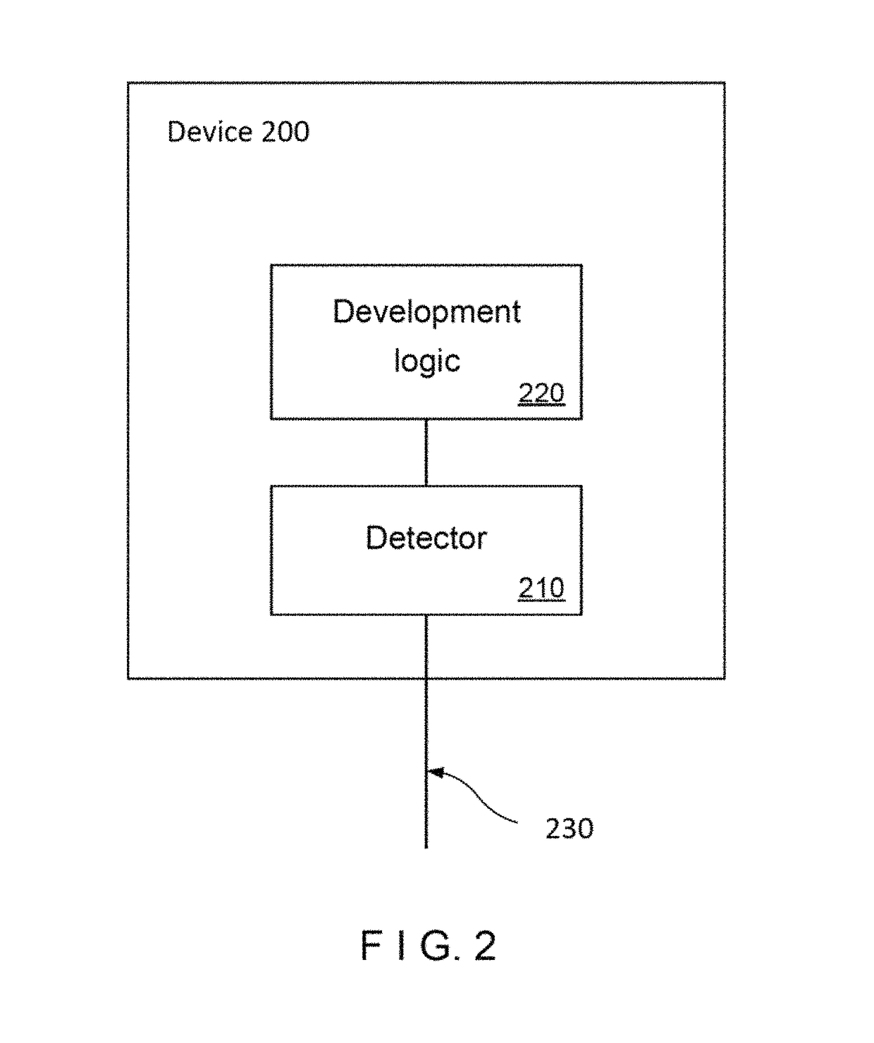
[0010]According to an exemplary embodiment of the present disclosure, apparatus and method can be provided for generating information for at least one structure. For example, using at least one first arrangement, it is possible to receive at least one first radiation from the at least one structure and at least one second radiation from a reference, and interfere the first and second radiations to generate at least one third radiation. Further, with at least one second arrangement, it is possible to generate spectroscopic data as a function of the at least one third radiation, and reduce at least one scattering effect in the spectroscopic data to generate the information.
[0011]For example, the second arrangement(s) can be used to reduce an intensity variation of the of the spectroscopic data. The second arrangement(s) can further be used to smooth the spectroscopic data based a polynomial fit to the spectroscopic data. The structure(s) can be one or more anatomical structures. The anatomical structure(s) can be an artery wall. The second arrangement(s) can be further used to classify a type of the structure based on the spectroscopic data.

Problems solved by technology

While OCT techniques has been demonstrated to be capable of visualizing lipid pools, such lipid pools appear as the absence of OCT signal, making them possibly difficult to definitively identify.
Furthermore, OCT techniques can be subject to artifacts that give the appearance of lipid pool when one may not be present.
Beyond atherosclerosis, the interpretation of images in other organ systems, such as the GI tract, urinary tract, lung, breast, brain, head and neck can also be challenging as the conventional mode of contrast in OCT is scattering and does not directly provide information on the chemical and molecular composition of the tissue.

Method used


Image

  • Apparatus, systems, methods and computer-accessible medium for spectral analysis of optical coherence tomography images
  • Apparatus, systems, methods and computer-accessible medium for spectral analysis of optical coherence tomography images
  • Apparatus, systems, methods and computer-accessible medium for spectral analysis of optical coherence tomography images

Examples


Embodiment Construction

[0008]It is therefore one of the objects of the present disclosure to reduce or address the deficiencies and / or limitations of such prior art approaches, procedures, methods, systems, apparatus and computer-accessible medium.

[0009]Herein, exemplary embodiments of systems, apparatus, methods and computer-accessible medium are described for depth resolved spectral analysis of OCT interferometric data and visualization methods for the identification of lipid rich regions. According to one exemplary embodiment, an analysis of inteferometric signals acquired (or previously acquired) with current coronary OCT catheters and systems can be utilized for spatial identification of lipid rich regions. The combined exemplary architectural and chemical analysis can improve contrast of intracoronary OCT images, which can increase the accuracy and time to which a user identifies diseased areas.

[0010]According to an exemplary embodiment of the present disclosure, apparatus and method can be provided...


PUM

PropertyMeasurementUnit
wavelengthsaaaaaaaaaa

Abstract

According to an exemplary embodiment of the present disclosure, apparatus and method can be provided for generating information for at least one structure. For example, using at least one first arrangement, it is possible to receive at least one first radiation from the at least one structure and at least one second radiation from a reference, and interfere the first and second radiations to generate at least one third radiation. Further, with at least one second arrangement, it is possible to generate spectroscopic data as a function of the at least one third radiation, and reduce at least one scattering effect in the spectroscopic data to generate the information. In addition or as an alternative, according to a further exemplary embodiment of the present disclosure, it is possible to classify a type of the structure based on the spectroscopic data to generate the information.

Description

CROSS-REFERENCE TO RELATED APPLICATION(S)[0001]The present application is based on and claims priority from U.S. Patent Application Ser. No. 61 / 348,108, filed on May 25, 2010, and U.S. Patent Application Ser. No. 61 / 406,494, filed on Oct. 25, 2010, the entire disclosures of which are incorporated herein by reference.STATEMENT OF FEDERALLY-SPONSORED RESEARCH AND DEVELOPMENT[0002]This invention was made with Government support under grant numbers HL076398 and HL000093 awarded by the National Institutes of Health. The Government has certain rights in this disclosure.FIELD OF THE DISCLOSURE[0003]Exemplary embodiments of the present disclosure relates generally to Spectral analysis of optical coherence tomography images, and more particularly to apparatus, systems, methods and computer accessible medium for an identification of components related to atherosclerotic plaque from OCT images through depth resolved spectral analysis of optical coherence tomography (OCT) images, including time...

Claims


Application Information

Patent Timeline
Patent Type & Authority Patents(United States)
IPC IPC(8): G01B9/02A61B5/00G01N21/47A61B5/02G01J3/45G01N21/17
CPCA61B5/0066A61B5/02007G01B9/02091G01N21/4795G01N2201/1293A61B5/7264G01N2021/1787G01N2201/129
Inventor FLEMING, CHRISTINEBOUMA, BRETT EUGENEGARDECKI, JOSEPHTEARNEY, GUILLERMO J.
Owner THE GENERAL HOSPITAL CORP
Learn More