Apparatus for finding a functional tissue area in a tissue region

a functional tissue and apparatus technology, applied in the field of apparatus for finding a functional tissue area in a tissue region, can solve the problem that the requirement of expensive monochrome cameras with associated color filters is not mandatory, and achieve the effect of enabling contrast-rich imaging of the functional tissue area in a short period of time and cost-effectiveness

Active Publication Date: 2017-10-31
TECHN UNIV DRESDEN INST FUER BIOMEDIZINISCHE TECHN +2
View PDF6 Cites 9 Cited by
  • Summary
  • Abstract
  • Description
  • Claims
  • Application Information

AI Technical Summary

Benefits of technology

[0007]It is an object of the invention to develop and provide an apparatus for finding a functional tissue area in a tissue region. The apparatus is cost-effective and enables contrast-rich imaging of the functional tissue area in a short period of time. It is furthermore an object of the invention to develop and provide a method for finding a functional tissue area in a tissue region. It is furthermore an object of the invention to develop a surgical microscope comprising such an apparatus.
[0015]Preferably, the at least one optical property is the intensity or wavelength of the reflected light. A stimulation of the tissue region leads to neuronal activity and therefore to an increased oxygen consumption. This results in an expansion of arterioles and therefore to higher blood flow with a higher blood volume in the corresponding tissue region. In addition to such a change in the blood volume, there moreover is a change in the oxygen saturation of the hemoglobin in the blood. The increased oxygen requirement brings about an increase in the deoxygenated hemoglobin and oxygenated hemoglobin. If such a tissue region is illuminated, this leads to a change in the absorption of the reflected light. Thus, this is therefore accompanied by a change in the intensity of the reflected light, wherein the change in intensity can be acquired by a camera. If the camera acquires such a change in the intensity in a wavelength range between 400 nm and 520 nm and / or in a wavelength range between 470 nm and 600 nm, this is enough to identify a functional tissue area in the tissue region.
[0016]Preferably, the change in the at least one optical property is pulsed. This can be achieved by virtue of the stimulation being brought about in a pulsed manner. Thus, there is a periodic change between the cortical stimulation and no stimulation, which is easily implementable by means of process control.
[0023]Using this method, a contrast-rich image of a functional tissue area in a tissue region can be obtained in a cost-effective and quick manner, and with little complexity.

Problems solved by technology

The inventors have identified that an expensive monochrome camera with an associated color filter is not mandatory for being able to find a functional tissue area in a tissue region.

Method used


Image

  • Apparatus for finding a functional tissue area in a tissue region
  • Apparatus for finding a functional tissue area in a tissue region
  • Apparatus for finding a functional tissue area in a tissue region

Examples


Embodiment Construction

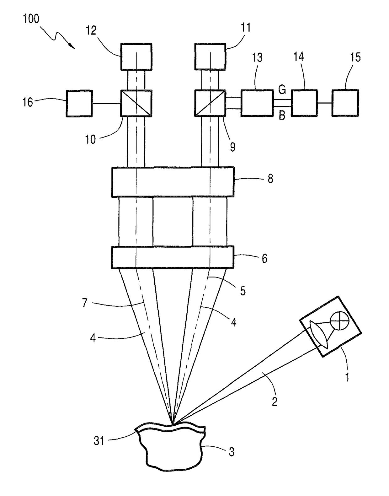
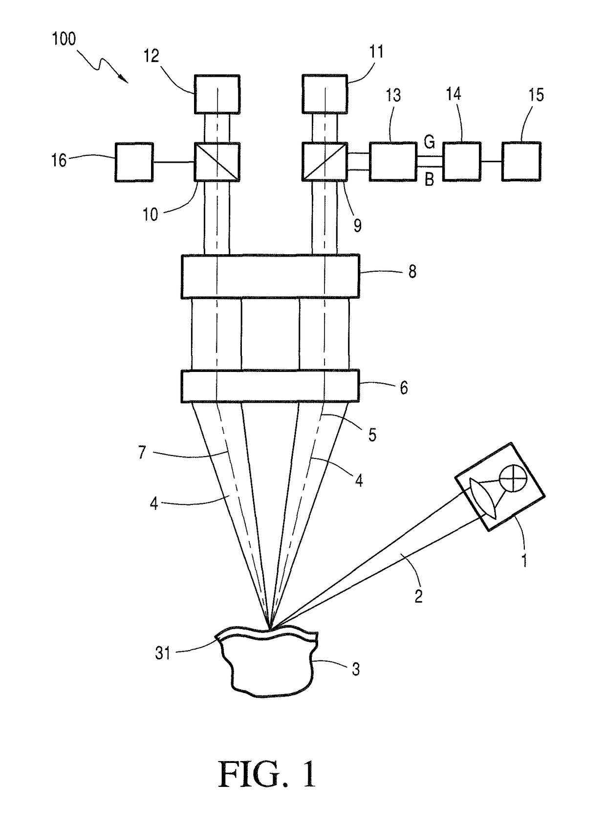
[0028]FIG. 1 schematically depicts an apparatus 100 according to the invention in accordance with a first embodiment for finding a functional tissue area in a tissue region 3. The apparatus 100 has a measurement illuminating device 1 which may have a xenon lamp or halogen lamp. The measurement illuminating device 1 transmits white light 2 to a tissue area in a tissue region 3 which, for example, has a blood path 31. The light 4 reflected by the blood path 31 reaches an objective 6 in a first beam path 5 and, from there, it reaches a magnification changer 8. From there, the light reaches a first beam splitter 9, from where the light reaches an eyepiece 11 and a camera 13. The camera 13 has a green channel G and a blue channel B, which are both coupled to an evaluation unit 14. When the tissue area in the tissue region 3 is stimulated in such a way that there is a change in the concentration of oxygenated and deoxygenated hemoglobin, this brings about a change in the absorption of the...


PUM


Abstract

The invention is directed to an apparatus for finding a functional tissue area in a tissue region. The apparatus has a measurement illuminating device suitable for emitting measurement illumination to the tissue region and a camera which can capture light reflected by the tissue region. The camera has a green channel and / or a blue channel wherein there is a change in an optical property of the light reflected by the tissue region during the stimulation thereof which is undertaken at least intermittently. An evaluation unit captures the change in the optical property only by a signal of the green channel and / or of the blue channel of the camera. A display unit can display an output signal of the evaluation unit for the functional tissue area in the tissue region.

Description

CROSS REFERENCE TO RELATED APPLICATION[0001]This application claims priority of German patent application no. 10 2014 005 407.1, filed Apr. 14, 2014, the entire content of which is incorporated herein by reference.FIELD OF THE INVENTION[0002]The present invention relates to an apparatus for finding a functional tissue area in a tissue region, especially in a brain tissue region. The invention further relates to a method for finding a functional tissue area in a tissue region and a surgical microscope having an apparatus referred to above.BACKGROUND OF THE INVENTION[0003]In the case of tumor surgery in the brain with a tumor in the vicinity of eloquent brain areas, that is, functional brain areas, such as, for example, in the vicinity of the motor cortex, in the vicinity of the sensorimotor centers or in the vicinity of the speech center, a surgeon is presented with the conflicting goals of, on the one hand, radically removing the tumor and, on the other hand, removing as little tiss...

Claims


Application Information

Patent Timeline
Patent Type & Authority Patents(United States)
IPC IPC(8): A61B5/00A61B5/026A61B5/1455A61B90/20
CPCA61B5/0075A61B5/0261A61B5/14553A61B90/20A61B5/742A61B2576/02A61B2505/05G16H30/40
Inventor PANITZ, GERALDHAUGER, CHRISTOPHWILZBACH, MARCOGUCKLER, ROLANDMEYER, TOBIASOELSCHLAEGEL, MARTINMORGENSTERN, UTESOBOTTKA, STEPHAN B.SCHACKERT, GABRIELE
Owner TECHN UNIV DRESDEN INST FUER BIOMEDIZINISCHE TECHN
Why Eureka
Try Eureka