
Multimode X-ray apparatus and method for the operation

a multi-mode x-ray and operation technology, applied in the field of multi-mode x-ray apparatus and operation, can solve the problems of increasing increasing the dose of anti-scattering grids, and increasing the quality of image contrast and hence image quality, so as to shorten the recording procedure and reduce the manufacturing cost of the multi-mode x-ray device. the effect of reliability and shortening the recording procedur

Active Publication Date: 2017-12-05
SIEMENS HEALTHCARE GMBH
View PDF8 Cites 0 Cited by
  • Summary
  • Abstract
  • Description
  • Claims
  • Application Information

AI Technical Summary

Benefits of technology

This approach reduces radiation exposure by up to 30% and eliminates potential sources of image artifacts, improving patient comfort and device reliability, while maintaining high-quality image production without the need for an anti-scatter grid.

Problems solved by technology

The scattered radiation causes more image noise and reduces the quality of the image contrast and hence also the image quality.
The problem of the dose being increased by the use of an anti-scatter-grid also exists in the multimode x-ray devices known in the prior art because, in that case, an anti-scatter grid is also situated in the beam path in order to absorb the scattered radiation when producing the mammogram.

Method used


Image

  • Multimode X-ray apparatus and method for the operation
  • Multimode X-ray apparatus and method for the operation
  • Multimode X-ray apparatus and method for the operation

Examples


Embodiment Construction

[0027]All figures show the invention merely schematically and with the basic components thereof. The same reference signs correspond to elements with the same or a comparable function.

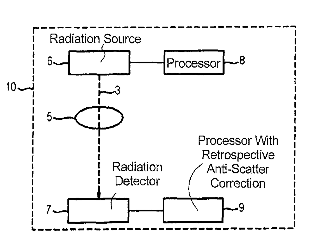
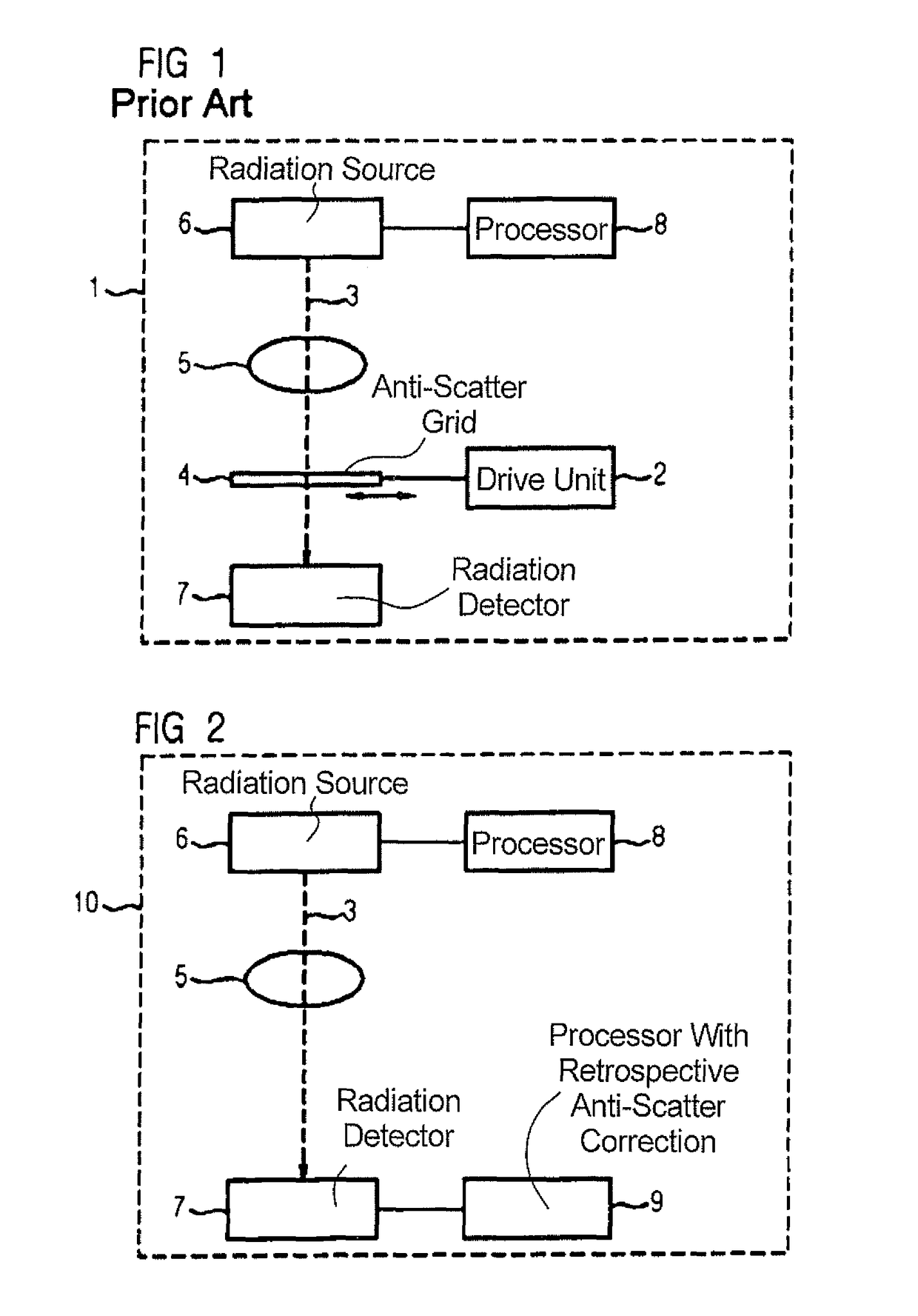
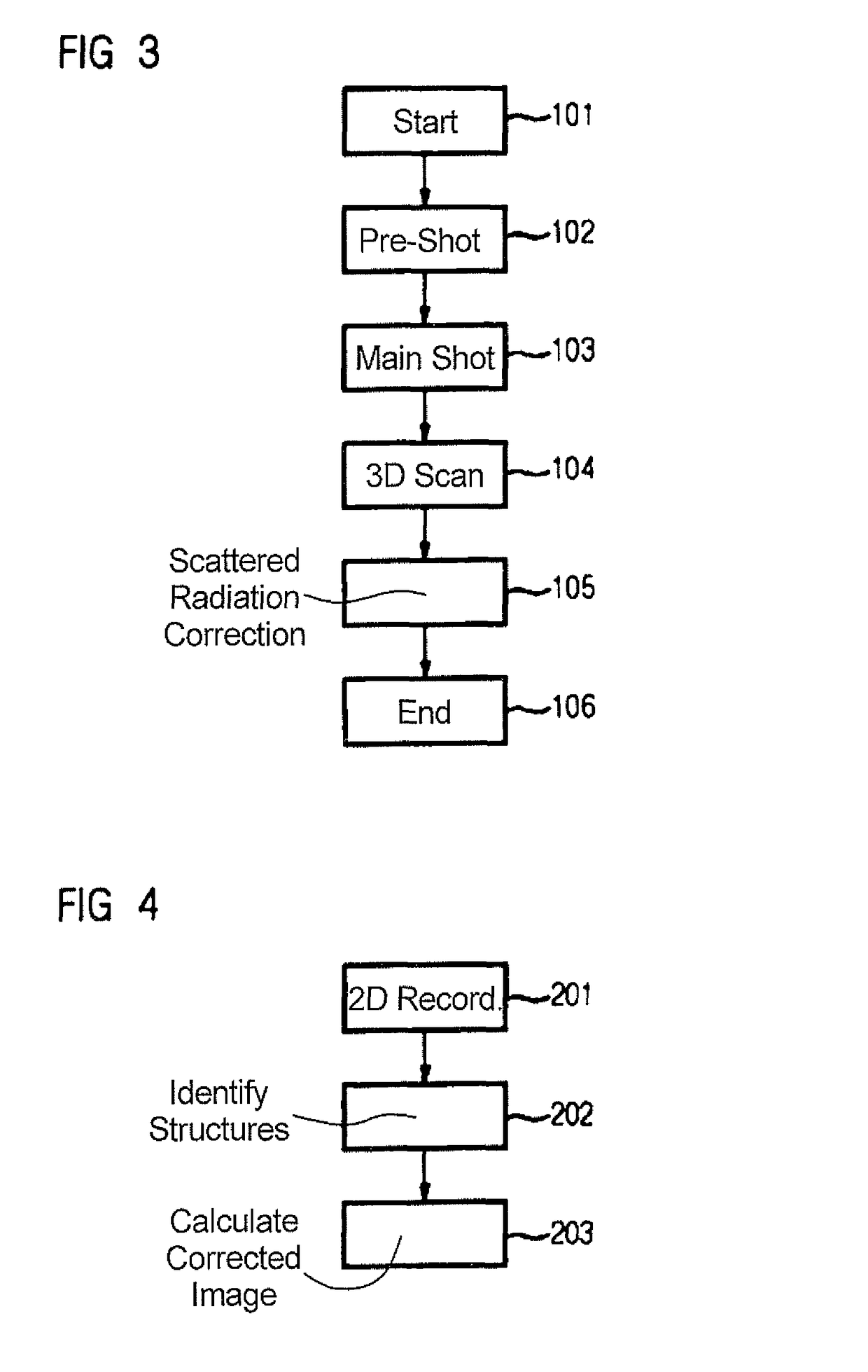
[0028]FIG. 1 shows a multimode x-ray device 1, as is known from the prior art, that has an anti-scatter grid 4, that is insertable into the beam path 3 with the use of a drive unit 2, when required. The anti-scatter grid 4 and drive unit 2 are dispensed within the multimode x-ray device 10 according to the invention, as is depicted in FIG. 2. The multimode x-ray device 10 is operable in two recording modes. It is embodied to produce 2-D recordings and 3-D recordings of a breast 5 of a patient. It has an x-ray radiation source 6 and an x-ray radiation detector 7. The breast 5 is placed between the x-ray radiation source 6 and the x-ray radiation detector 7. The 2-D recordings and the 3-D recordings are producible with the same breast placement.

[0029]As depicted in FIG. 3, there initially is a grid-free ...


PUM


Abstract

In a method for producing 2-D recordings and 3-D recordings of a breast of a patient using an x-ray device that is operable in two recording modes and that has an x-ray radiation source and an x-ray radiation detector, the breast is placed between the x-ray radiation source and the x-ray radiation detector, and the 2-D recordings and the 3-D recordings are generated with the same breast placement. In order to keep the radiation exposure as low as possible in such a multi-mode x-ray device, all recordings are produced without the use of an anti-scatter grid, and a retroactive scattered radiation correction is implemented.

Description

BACKGROUND OF THE INVENTION[0001]Field of the Invention[0002]The invention concerns a method for producing 2-D recordings and 3-D recordings of a breast of a patient using an x-ray apparatus of the type that is operable in two recording modes and that has an x-ray radiation source and an x-ray radiation detector, wherein the breast is placed between the x-ray radiation source and the x-ray radiation detector, wherein the 2-D recordings and the 3-D recordings are generated with the same breast placement.[0003]Moreover, the invention concerns an x-ray apparatus that is operable in two recording modes, designed for producing 2-D recordings and 3-D recordings of a breast of a patient, of the type having an x-ray radiation source and an x-ray radiation detector, wherein the breast is placeable between the x-ray radiation source and the x-ray radiation detector, wherein the 2-D recordings and 3-D recordings are producible with the same breast placement.[0004]Description of the Prior Art[0...

Claims


Application Information

Patent Timeline
Patent Type & Authority Patents(United States)
IPC IPC(8): A61B6/00G06T11/00G06T5/00
CPCG06T11/005A61B6/466A61B6/502A61B6/5205A61B6/5282A61B6/54G06T5/002G06T2211/421A61B6/563G06T2207/10081G06T2207/10116G06T2207/30068G06T2210/41G06T11/008G06T2211/436G06T5/70
Inventor HOERNIG, MATHIASMERTELMEIER, THOMAS
Owner SIEMENS HEALTHCARE GMBH
Learn More