Segmentation apparatus for interactively segmenting blood vessels in angiographic image data

a segmentation apparatus and angiographic image data technology, applied in image enhancement, image analysis, instruments, etc., can solve the problems of missing intermediate-grade blood vessels and time-consuming segmentation procedures, and achieve the effect of easing the burden on the operator

Active Publication Date: 2017-12-12
KONINKLJIJKE PHILIPS NV
View PDF34 Cites 12 Cited by
  • Summary
  • Abstract
  • Description
  • Claims
  • Application Information

AI Technical Summary

Benefits of technology

[0006]It is an object of the present invention to provide a segmentation apparatus, a segmentation method, and a segmentation computer program for interactively segmenting blood vessels in angiographic image data, which allow to ease the burden on an operator during the segmentation procedure. It is a further object of the present invention to provide a system for calculating values of a blood flow parameter based on a segmentation of blood vessels in angiographic image data, wherein the system comprises the segmentation apparatus.
[0010]Since the significant location determining unit is adapted to determine one or more locations of a current segmentation of the blood vessels in the angiographic image data as significant locations, i.e., as locations at which the current segmentation has a predetermined influence on a value of a blood flow parameter that is calculated based on the current segmentation, and since the display unit is adapted to display the significant locations to an operator, the operator can be guided to focus his / her segmentation efforts to those locations of the current segmentation that are most relevant for the accuracy of the calculation of the value of the blood flow parameter. This allows to ease the burden on the operator during the segmentation procedure, since he / she may now only need to perform the segmentation with a higher degree of accuracy and, thus, a higher effort at the significant locations. In effect, this may result in a higher-quality segmentation of the blood vessels in the angiographc image data and, therewith, a higher-accuracy calculation of values of the blood flow parameter based on the segmentation.
[0016]In an embodiment, the criterion is based on a rate of change of the diameter of the current segmentation at the one or more locations. For instance, a location of the current segmentation can be determined as a significant location candidate if the rate of change which may, e.g., be calculated as a simple derivative in case the segmentation is represented by one or more mathematical functions, such as spline curves or straight segments interpolating a number of nodes of the segmentation of the diameter of the current segmentation at that location is large. This can be determined, e.g., by comparing the rate of change with a predefined threshold. Since the rate of change of the diameter of the current segmentation can be calculated in a simple manner, and since e.g. stenoses, which in general have a significant influence on the blood flow in the blood vessels, are typically characterized by rapid changes in blood vessel diameter, the rate of change of the diameter of the current segmentation can be a good criterion for determining the significant location candidates.
[0019]It is preferred that the display unit is adapted to display the current segmentation with the angiographic image data to the operator, wherein the significant locations are indicated by means of visual indications. This provides an easy and efficient way of guiding the operator to focus his / her segmentation efforts to the significant locations. The visual indications can comprise, e.g., signs or other visual marks, such as boxes, crosses, triangles, circles, pointers, stippled lines, or the like, that indicate the significant locations of the current segmentation. Additionally or alternatively, color codings, specific shadings, or the like, can be used as visual indications.
[0024]It is preferred that the segmentation apparatus comprises a segmentation modifying unit for allowing the operator to modify the current segmentation at least at the significant locations. This allows the operator to update and improve the current segmentation at those locations that are most relevant for the accuracy of the calculation of the value of blood flow parameter. The modification can be, e.g., fully manual or semi-automatic. For example, the operator can be able to add or remove one or more nodes for refining the current segmentation at the significant locations. Of course, the segmentation modifying unit can also be adapted for allowing the operator to modify the current segmentation at locations other than the significant locations.

Problems solved by technology

The problem is that such segmentation can be a very time-consuming procedure, which typically involves semi-automatic image processing algorithms that require interactive control by an operator, e.g., a physician, in order to segment each branch of the coronary arterial tree.
In addition, operators often only rely on their visual assessment for deciding when the segmentation is “good enough”.
This may lead to the missing of intermediate-grade, but clinically relevant stenoses (i.e., abnormal narrowings or constrictions of the diameter of a blood vessel) in the coronary arteries.

Method used


Image

  • Segmentation apparatus for interactively segmenting blood vessels in angiographic image data
  • Segmentation apparatus for interactively segmenting blood vessels in angiographic image data
  • Segmentation apparatus for interactively segmenting blood vessels in angiographic image data

Examples


Embodiment Construction

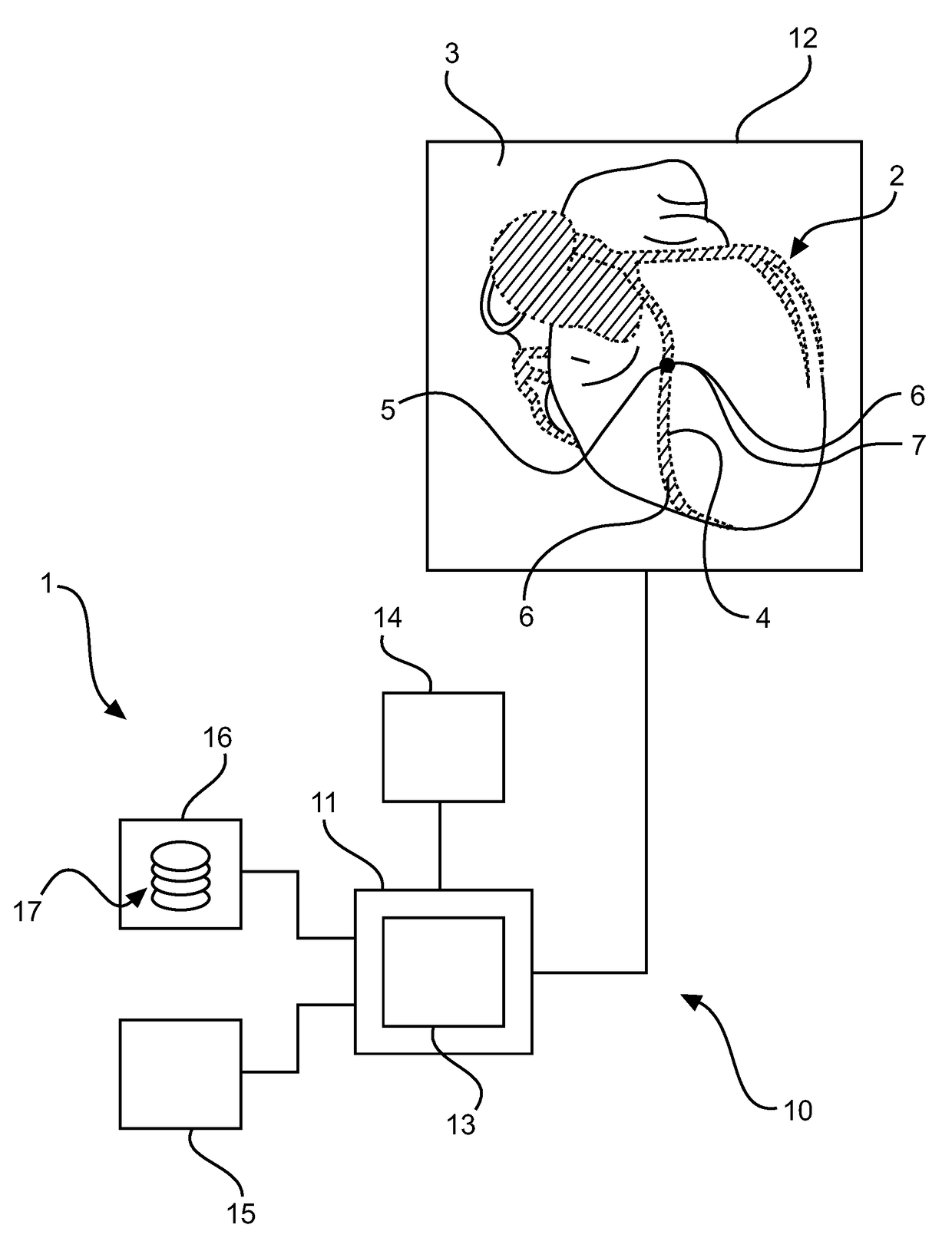
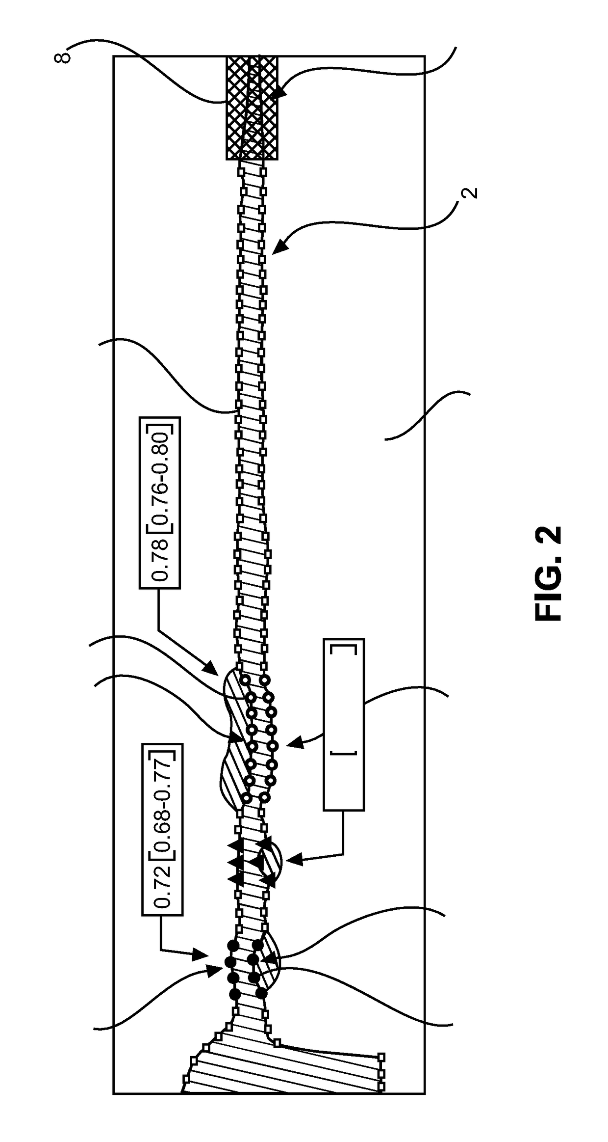
[0040]FIG. 1 shows schematically and exemplarily an embodiment of a system 1 for calculating values of a blood flow parameter, here, a virtual fractional flow reserve (vFFR), based on a segmentation 4 of blood vessels 2, in this example, coronary arteries of a human being (not shown in the figure), in angiographic image data 3. The system 1 comprises a segmentation apparatus 10 for interactively segmenting the coronary arteries 2 in the angiographic image data 3, and a blood flow parameter calculation unit 14 for calculating the values of the vFFR.

[0041]The segmentation apparatus 10 comprises a significant location determining unit 11 for determining one or more locations of a current segmentation 4 of the coronary arteries 2 in the angiographic image data 3 as significant locations 5, i.e., as locations at which the current segmentation 4 has a predetermined influence on a value of the vFFR that is calculated based on the current segmentation 4, and a display unit 12 for displaying...


PUM


Abstract

The present invention relates a segmentation apparatus (10) for interactively segmenting blood vessels (2) in angiographic image data (3). The segmentation apparatus (10) comprises a significant location determining unit (11) for determining one or more locations of a current segmentation (4) of the blood vessels (2) in the angiographic image data (3) as significant locations (5) at which the current segmentation (4) has a predetermined influence on a value of a blood flow parameter that is calculated based on the current segmentation (4), and a display unit (12) for displaying the significant locations (5) to an operator. Thereby, the operator can be guided to focus his / her segmentation efforts to those locations of the current segmentation (4) that are most relevant for the accuracy of the calculation of the value of the blood flow parameter. This may ease the burden on the operator during the segmentation procedure.

Description

CROSS REFERENCE TO RELATED APPLICATIONS[0001]This application is the U.S. National Phase application under 35 U.S.C. §371 of International Application No. PCT / EP2014 / 067660, filed Aug. 19, 2014, published as WO 2015 / 024934 on Feb. 26, 2015, which claims the benefit of European Patent Application Number 13181139.0 filed Aug. 21, 2013. These applications are hereby incorporated by reference herein.FIELD OF THE INVENTION[0002]The present invention relates to a segmentation apparatus, a segmentation method, and a segmentation computer program for interactively segmenting blood vessels in angiographic image data. The present invention relates further to a system for calculating values of a blood flow parameter based on a segmentation of blood vessels in angiographic image data, wherein the system comprises the segmentation apparatus.BACKGROUND OF THE INVENTION[0003]US 2011 / 0071404 A1 discloses a computer-based system for evaluating a region of a lumen. The system comprises an image acqui...

Claims


Application Information

Patent Timeline
Patent Type & Authority Patents(United States)
IPC IPC(8): G06K9/00G06T7/00G06T7/11A61B5/02
CPCG06T7/11G06T7/0012G06T2207/20104G06T2207/30104G06T2210/41G06T2207/30048
Inventor PREVRHAL, SVENNICKISCH, HANNESSCHMITT, HOLGER
Owner KONINKLJIJKE PHILIPS NV
Why Eureka
Try Eureka