Treatment to promote wound healing
a technology for wound healing and treatment, applied in the field of tissue repair and regeneration, can solve the problems of serious compromising host fitness, poor school or work performance, etc., and achieve the effect of promoting or accelerating wound healing
- Summary
- Abstract
- Description
- Claims
- Application Information
AI Technical Summary
Benefits of technology
Problems solved by technology
Method used
Image
Examples
example 1
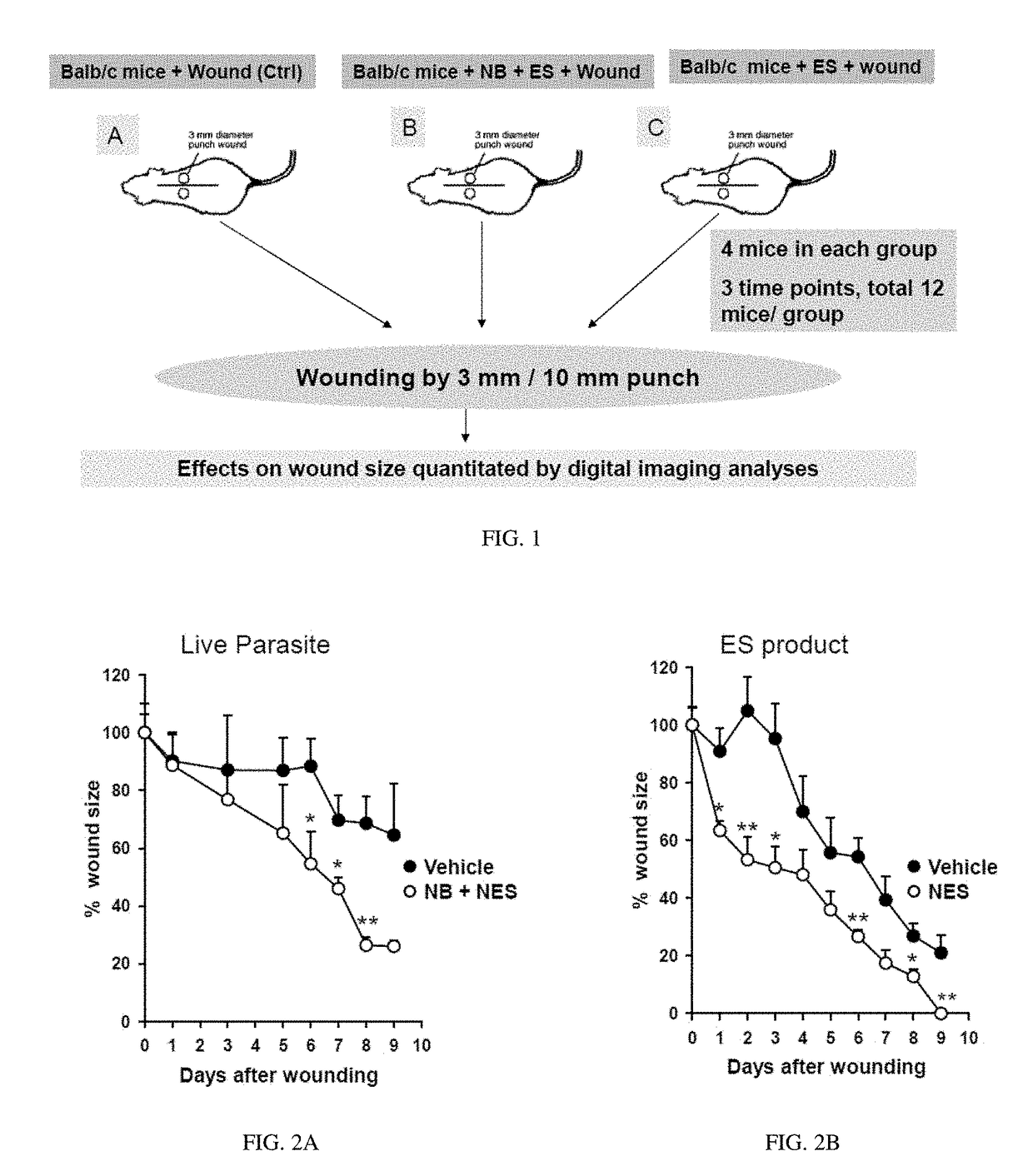
[0059]To examine whether helminthes accelerate cutaneous wound healing, punch biopsies were used to create defined cutaneous wounds in mice based on a protocol previously described (Chigurupati, S., et al. PLoS One 5, e10044 (2010); Reiss, M. J., et al. Surgery 147, 295-302 (2010); Syeda, M. M., et al. Am J Pathol 181, 334-346 (2012); Ojha, N., et al. Free Radic Biol Med 44, 682-691 (2008); incorporated herein by reference in their entirety). Specifically, C57BL.6 male mice, age 8-10 weeks, were used for this model experiment. The backs of the mice were shaved with standard animal hair clippers and disinfected with betadine (Fisher, N.J., USA). After 48 hours, mice were infected either with 500 Nippostrongylus brasiliensis (Nb) third-stage larvae (L3) or left untreated (control). 24 hours after the Nb infection, 50 microgram of NES product derived from both Nb L3 larvae and Nb L5 adult worms were injected subcutaneously at the site of wounding. Forty-eight hours after Nb inoculation...
example 2
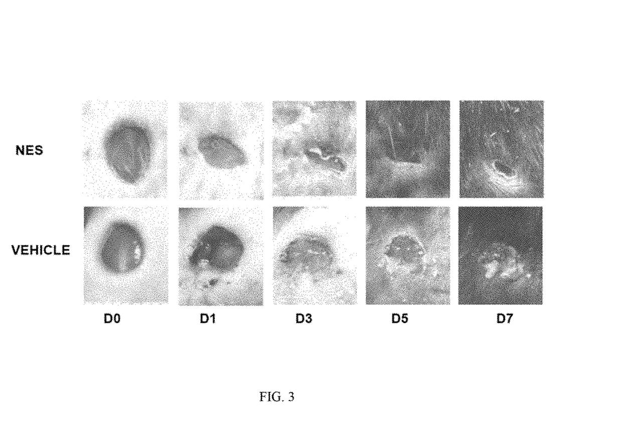
[0060]In order to assess the potential of parasite and parasite derived products, NES was collected form L3 and L5 larvae, concentrated (as described in Ojha, 2008), and analyzed for contaminating endotoxin present in the NES by using an endpoint chromomeric Limulus amebocyte lysate assay (QCL-1000; Lonza, Walkersville, Md.). The LPS levels of NES products were found to be less than 3 units 1 ml of NES preparation. Mice were infected with 300 L3 and administered NES subcutaneously 24 later. Forty-eight hours post infection 5 mm punch biopsies were administered to either naïve mice or mice treated with Nb+NES. Three days after wounding, 50 micrograms of NES was applied over the wound. Wound closure was assessed over a period of 9 days and digital photographs of wound closure taken. Using digital imaging analysis, the area of the wound was quantitated in controls and in mice administered Nb+NES. As shown in FIG. 2A, a statistically significant enhanced wound closure was found to occur...
example 3
[0061]An experimental in vivo wound healing experiments shown in Examples 1 and 2 has been repeated with NES alone. Specifically. NES was subcutaneously injected at the site of punch biopsy 24 hours before treatment. NES was then administered via subcutaneous injection every 24 hours thereafter. Biopsies were photographed on a daily basis and the wound area quantitated as previously described. In these studies, a dose response experiment reveals that administration of NES alone is sufficient to accelerate wound closure as shown in FIG. 2B.
[0062]Tissues were collected at specific timepoints and assess through immunohistological techniques changes in inflammatory cells, the degree of fibrosis, reepithelialization, angiogenesis, and tissue granulation. Tissues were also collected to examine changes in gene expression with a particular focus on genes associated with wound healing and the helminth-induced type 2 immune response including: specific Th1 and Th2 cytokines, collagens, colleg...
PUM
| Property | Measurement | Unit |
|---|---|---|
| fecundity energy consumption | aaaaa | aaaaa |
| parasite resistance | aaaaa | aaaaa |
| time | aaaaa | aaaaa |
Abstract
Description
Claims
Application Information
 - R&D
- Intellectual Property
- Life Sciences
- Materials
- Tech Scout
- Unparalleled Data Quality
- Higher Quality Content
- 60% Fewer Hallucinations
Browse by: Latest US Patents, China's latest patents, Technical Efficacy Thesaurus, Application Domain, Technology Topic, Popular Technical Reports.
© 2025 PatSnap. All rights reserved.Legal|Privacy policy|Modern Slavery Act Transparency Statement|Sitemap|About US| Contact US: help@patsnap.com


