
System and method for multi-axis imaging of specimens

a multi-axis imaging and specimen technology, applied in the field of tissue specimen analysis, can solve the problems of inaccurate diagnosis, difficult or even impossible accurate tissue margin analysis, etc., and achieve the effects of facilitating accurate and efficient multi-axis imaging, diagnosis, and facilitating substantially horizontal or flat imaging

Active Publication Date: 2018-04-17
FAXITRON BIOPTICS
View PDF5 Cites 4 Cited by
  • Summary
  • Abstract
  • Description
  • Claims
  • Application Information

AI Technical Summary

Benefits of technology

The solution ensures accurate and efficient multi-axis imaging, maintains specimen integrity during transport, and improves diagnostic accuracy by maintaining the specimen's stability and reducing image artifacts.

Problems solved by technology

For instance, reshaping of the tissue specimen (e.g., compressing, folding, etc.) between the taking of first and second orthogonal images (e.g., for use in tissue margin detection) can make accurate tissue margin analysis difficult or even impossible.
As another example, the pathologist may have difficulty reconciling the locations on the resulting image identified by the surgeon or radiologist and corresponding locations on the actual excised tissue specimen and possibly leading to an inaccurate diagnosis in the event that the tissue specimen is moved relative to the grid or coordinate system during transport from the surgeon or radiologist to the pathologist.

Method used


Image

  • System and method for multi-axis imaging of specimens
  • System and method for multi-axis imaging of specimens
  • System and method for multi-axis imaging of specimens

Examples


Embodiment Construction

[0057]Reference will now be made to the accompanying drawings, which assist in illustrating the various pertinent features of the various novel aspects of the present disclosure. In this regard, the following description is presented for purposes of illustration and description. Furthermore, the description is not intended to limit the inventive aspects to the forms disclosed herein. Consequently, variations and modifications commensurate with the following teachings, and skill and knowledge of the relevant art, are within the scope of the present inventive aspects.

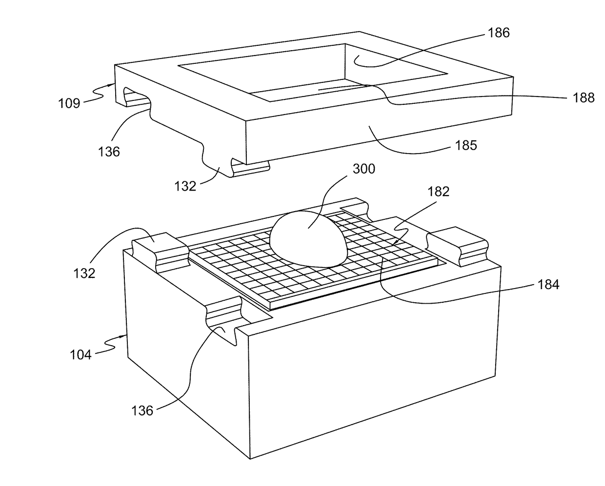
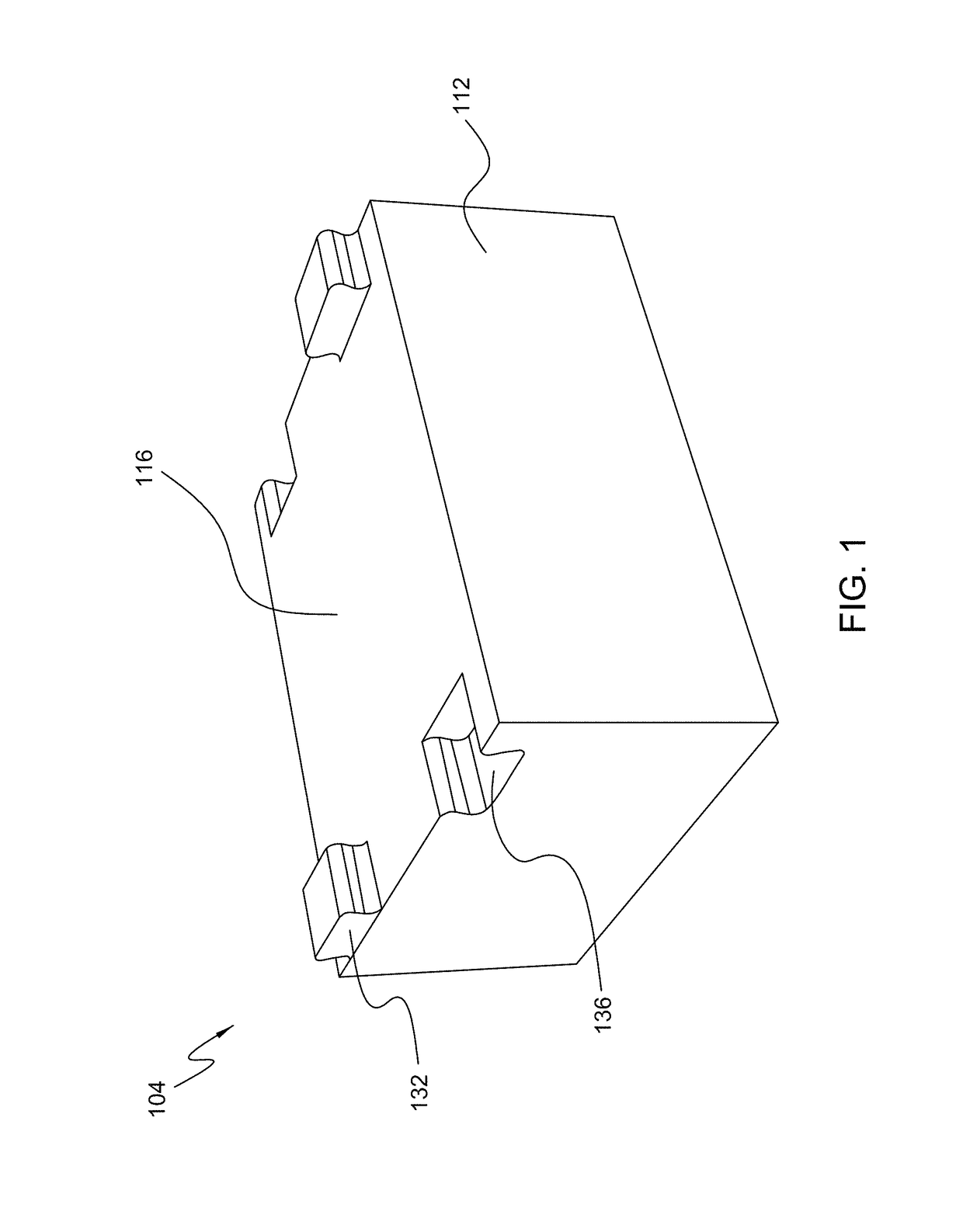
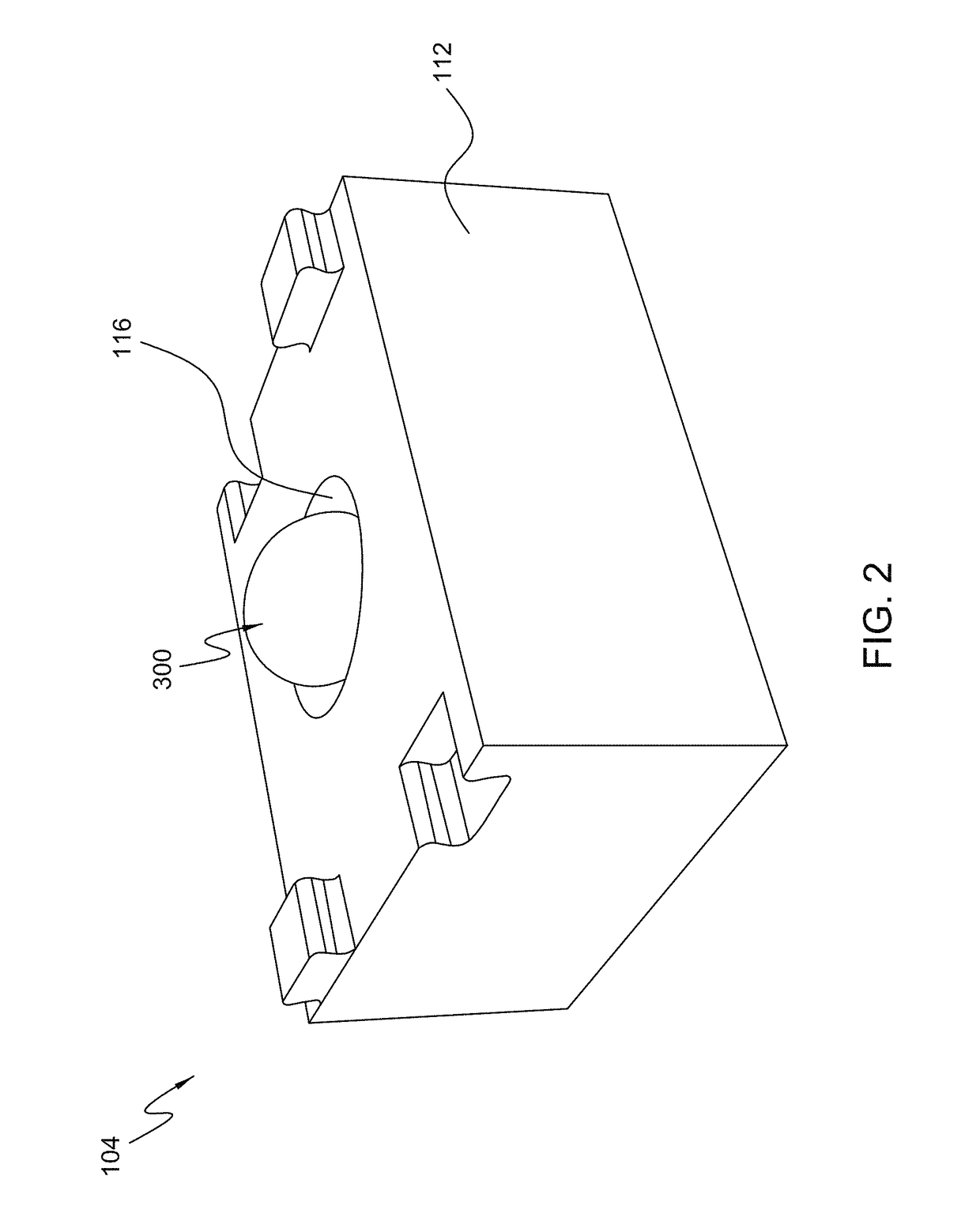
[0058]With initial respect to FIGS. 1-5, a specimen holding and positioning apparatus 100 is disclosed that is operable to maintain a specimen 300 (e.g., an excised tissue specimen) in a fixed or stable orientation with respect to the apparatus 100 during imaging operations (e.g., x-ray imaging), transport (e.g., from a surgery room to a pathologist's laboratory), and the like to facilitate the accurate detection and diag...


PUM

PropertyMeasurementUnit
volumeaaaaaaaaaa
densityaaaaaaaaaa
polymericaaaaaaaaaa

Abstract

A specimen holding and positioning apparatus operable to substantially non-movably maintain a specimen (e.g., an excised tissue specimen) in a fixed or stable orientation with respect to the apparatus during imaging operations (e.g., x-ray imaging), transport (e.g., from a surgery room to a pathologist's laboratory), and the like for use in facilitating accurate detection and diagnosis of cancers and / or other abnormalities of the specimen.

Description

CROSS REFERENCE TO RELATED APPLICATIONS[0001]This application is a continuation of International Patent App. No. US / PCT2015 / 017786, entitled “SYSTEM AND METHOD FOR MULTI-AXIS IMAGING OF SPECIMENS,” and filed on Feb. 26, 2015, which claims priority to U.S. Provisional Patent App. No. 62 / 045,073, entitled “SYSTEM AND METHOD FOR MULTI-AXIS IMAGING OF SPECIMENS,” and filed on Sep. 3, 2014, and to U.S. Provisional Patent App. No. 61 / 948,462, entitled “SYSTEM AND METHOD FOR MULTI-AXIS IMAGING OF SPECIMENS,” and filed on Mar. 5, 2014, the entire contents of which are incorporated herein by reference as if set forth in full.BACKGROUND OF THE INVENTION[0002]Field of the Invention[0003]This invention generally relates to tissue specimen analysis and, more particularly, to devices and methods for maintaining an excised tissue specimen in a fixed or stable orientation during imaging and transport to facilitate accurate detection of tissue margins, diagnosis of tissue abnormalities, and the like...

Claims


Application Information

Patent Timeline
Patent Type & Authority Patents(United States)
IPC IPC(8): G01N21/00B01L9/00G01N33/483A61B10/00
CPCB01L9/52G01N33/4833B01L2300/123B01L2300/0609B01L2300/0809A61B10/0096G01N2223/307G01N2223/6126
Inventor PURDY, CIARANO'DRISCOLL, DONOGH
Owner FAXITRON BIOPTICS
Learn More