
Knotless suture anchor assembly

a technology of suture anchor and assembly, which is applied in the direction of ligaments, surgical staples, prostheses, etc., can solve the problems of difficult and precise procedures, difficult and difficult use of bone tunnels for repair, and open incisions

Inactive Publication Date: 2003-01-07
THAL RAYMOND
View PDF35 Cites 397 Cited by
  • Summary
  • Abstract
  • Description
  • Claims
  • Application Information

AI Technical Summary

Benefits of technology

Further, the hollow anchoring sleeve can contain a collar on the rear portion or rear side of the hollow anchoring sleeve to control the depth of sleeve insertion into the bone and prevent excessive insertion depth.
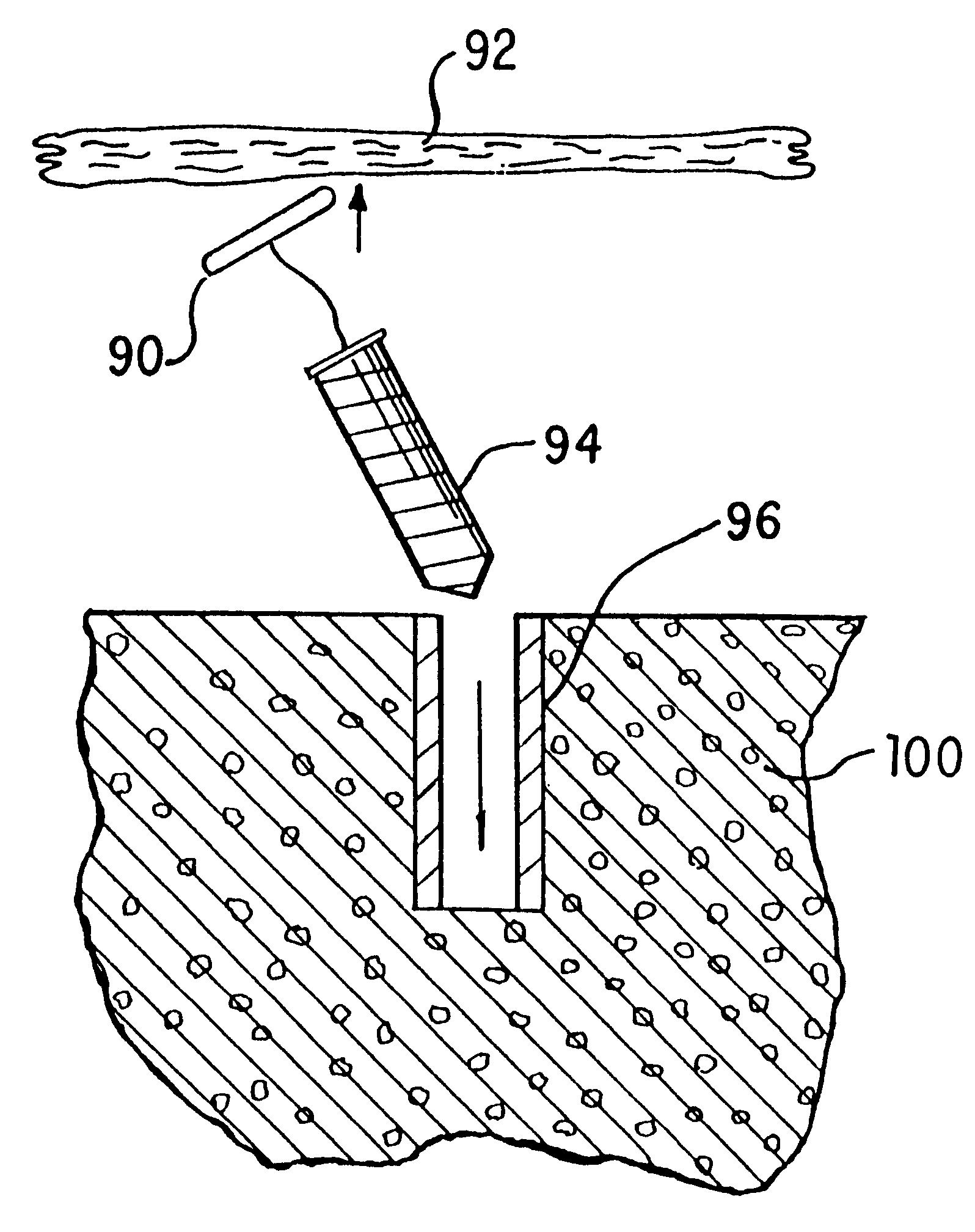
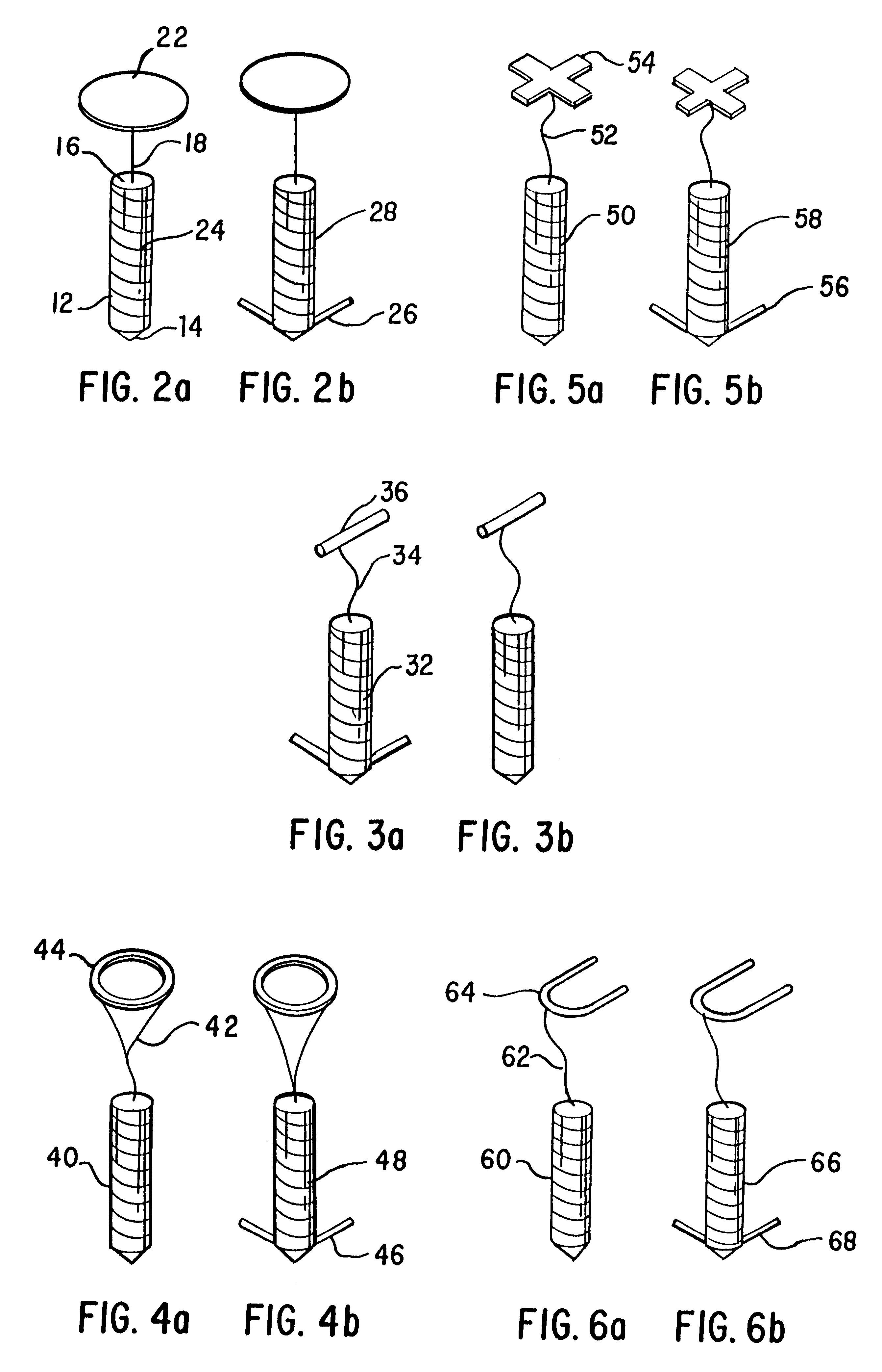
In the two-piece configuration, the spike or plug member is inserted during an open or endoscopic procedure, or the like, through the soft tissue and its piercing or pointed end is then inserted into the anchoring sleeve to facilitate a secure mating. Once the spike or plug member is threaded through the tissue and is inserted into the hollow anchoring sleeve, it is then securely attached through pressure by the surgeon into the sleeve. This attachment of the spike member to the hollow anchoring sleeve can be accomplished in one step or in a number of depth control steps (i.e., ratchets) to fine tune the tightness of the repair. This ratchet effect can be accomplished by a series of beads, ribs, thickening or the like on the exterior of the spike component. These would mate with the interior of the anchor sleeve. This allows for the tissue to be tightly attached to the bone mass. The unique device obviates the need for the surgeon to tie a knot with the suture material for reattachment of tissue to bone. Endoscopic procedures and some open surgical procedures are extremely difficult and must be completed in a very tight space. Obviation of the need of tying a knot is extremely beneficial and innovative.
The one-piece embodiment can be installed as described above. That is the spike or plug can be inserted through the soft tissue and then into the bone with the stop means grabbing the tissue and attaching or reattaching same to the bone mass. In the alternative, the stop means of the one-piece device can be inserted from the underside of the tissue mass through the mass, thereby obviating the need for the spike or plug with anchoring means from passing through the tissue. The spike or plug with anchor is then pressed into the bone mass pulling the tissue into attachment or reattachment to the bone mass.

Problems solved by technology

The use of bone tunnels for repair can be difficult and generally require large open incisions.
The unique knotless suture anchor assemblies described herein facilitate this difficult and precise procedure.
The process of passing the anchored suture through the soft tissue and tying a knot is time consuming and difficult to undertake in the tight space encountered during endoscopic surgery and sometimes even in conventional open surgery.
A few of these systems, such as those disclosed in U.S. Pat. Nos. 5,013,316 and 4,532,926, can be used arthroscopically but fixation with these devices may not be as secure as that achieved with sutures.
This type of non-suture staple device is disadvantageous in that it has been known to crack the bone during deployment, or accidentally transect the object being attached to the bone.
In addition, the device itself has been known to crack or break during or after deployment.

Method used


Image

  • Knotless suture anchor assembly
  • Knotless suture anchor assembly
  • Knotless suture anchor assembly

Examples


Embodiment Construction

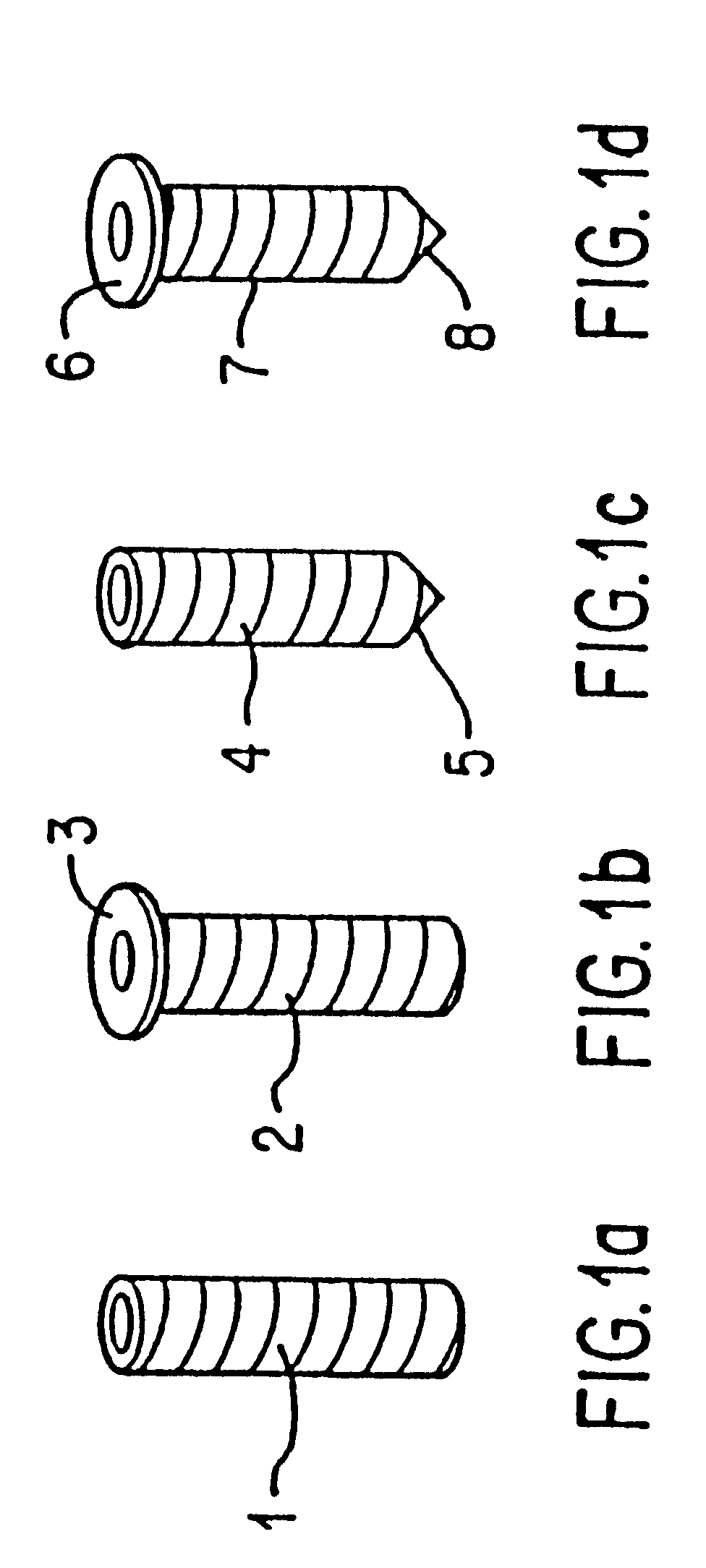
Referring now to FIG. 1, the knotless two-piece suture anchor assembly of the present invention contains as one integral component a hollow anchoring sleeve for installation and attachment to a bone mass. The hollow anchoring sleeve 1, as shown in FIG. 1a, is cylindrical in shape and possesses ribs or threads on its exterior. The device can also contain or be configured with prongs, umbrella spokes, have threads, be expandable, or have wedges, on its exterior, for secure attachment with the bone mass. These exterior attachment features are known to the industry and incorporated herein by reference.

FIG. 1b illustrates an alternate embodiment of the hollow anchoring sleeve 2 having a collar 3 to control depth of bone penetration. The collar prevents the sleeve from being forced too deep into the bone mass when the spike or plug member is inserted.

FIG. 1c illustrates an alternate embodiment of the hollow anchoring sleeve 4 wherein the sleeve has a pointed closed end 5 for ease of penet...


PUM


Abstract

A one-piece or two-piece knotless suture anchor assembly for the attachment or reattachment or repair of tissue to a bone mass. The assembly allows for an endoscopic or open surgical procedure to take place without the requirement of tying a knot for reattachment of tissue to bone mass. In one embodiment, a spike member is inserted through tissue and then inserted into a dowel-like hollow anchoring sleeve which has been inserted into a bone mass. The spike member is securely fastened or attached to the anchoring sleeve with a ratcheting mechanism thereby pulling or adhering (attaching) the tissue to the bone mass.

Description

BACKGROUND OF THE INVENTION1. Field of the InventionThe present invention relates to devices or assemblies used in tissue repair. More particularly, the assembly enables the attachment together or repair of portions of biological tissue (i.e., tendons or ligaments) onto a bone surface.2. Description of the Background ArtSoft tissues, such as tendons and ligaments, generally are attached to bone by small collagenous fibers. These connections are strong but permit the tendons and ligaments to be flexible. When a tissue is torn away from the bone and requires repair, a surgeon is often required to repair the detached soft tissue with sutures which are passed through bone tunnels and tied. A number of devices have been developed for securing a ligament or tendon to a bone mass. These devices can be used in place of bone tunnelling techniques. These attachment devices are usually inserted through extensive surgical incisions and, in some circumstances, by arthroscopic surgical techniques...

Claims


Application Information

Patent Timeline
IPC IPC(8): A61B17/04A61B17/56
CPCA61B17/0401A61F2/0811Y10T24/4698A61B2017/0403A61B2017/0404A61B2017/0412A61B2017/0414A61B2017/0417A61B2017/0419A61B2017/0427A61B2017/0437A61B2017/0438A61B2017/044A61F2002/0829A61F2002/0888Y10T24/3469Y10T24/4605Y10T24/4696A61B17/0487
Inventor THAL, RAYMOND
Owner THAL RAYMOND
Learn More