Methods for stable transduction of cells with viral vectors

A technology of lentiviral vectors and cells, which is applied in the direction of viruses/bacteriophages, viruses, and the use of vectors to introduce foreign genetic materials, etc., which can solve the problems that cells are not suitable for human clinical transplantation

Inactive Publication Date: 2008-12-17
VIRXSYS
View PDF5 Cites 2 Cited by
  • Summary
  • Abstract
  • Description
  • Claims
  • Application Information

AI Technical Summary

Problems solved by technology

Of note, use of this mixture would render these cells unsuitable for human clinical transplantation

Method used


Image

  • Methods for stable transduction of cells with viral vectors
  • Methods for stable transduction of cells with viral vectors
  • Methods for stable transduction of cells with viral vectors

Examples


Embodiment 1

[0107] Preparation of naive CD4+ T cells

[0108] With a slight modification of the standard method, peripheral blood CD4+ T cells were isolated. Specifically, confounding monocytes were removed using the adherence method. Non-adherent cells were contacted with magnetic beads coated with anti-CD4+ antibody to select for CD4+ cells. Take out the magnetic beads and separate the CD4+ cells.

[0109] The purity of highly purified CD4+ cells is as high as 90% through flow cytometry test.

Embodiment 2

[0111] Naive CD4+ T cells were transduced with cell surface binding molecules at different contact times.

[0112] Transduction prior to cell surface binding

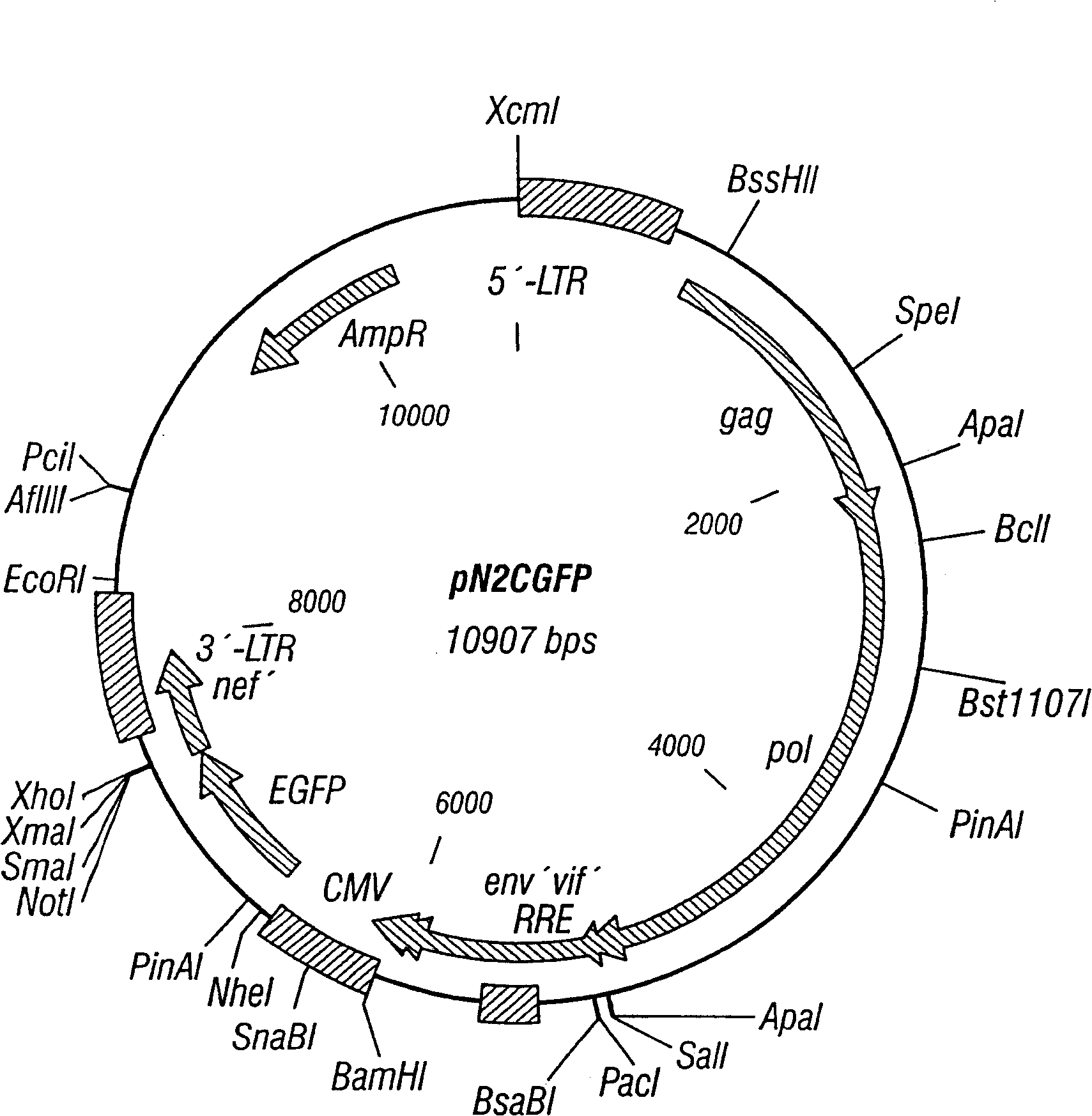
[0113] Naive CD4+ cells (about 500,000 cells) were co-cultured with pN2cGFP at MOI 20 for 24 hours, then added microbeads coated with αCD3 and αCD28, and cultured for another 7 days. Figure 1 shows the map of pN2cGFP. Transduction after cell surface binding

[0114] Naive CD4+ cells (approximately 500,000) were co-cultured with αCD3 and αCD28-coated microbeads for 24 hours, then pN2cGFP was added to the culture at an MOI of 20, and co-culture was continued for 24 hours. Then, they were cultured for 7 days in a medium containing microbeads but no carrier to wash away excess carrier.

[0115] Simultaneous cell surface binding and transduction

[0116] Naive CD4+ cells (~500,000) were co-cultured at MOI 20 with pN2cGFP for 24 hours in the presence of microbeads coated with αCD3 and αCD28. Then, they were cultured fo...

Embodiment III

[0121] Post-transduction analysis

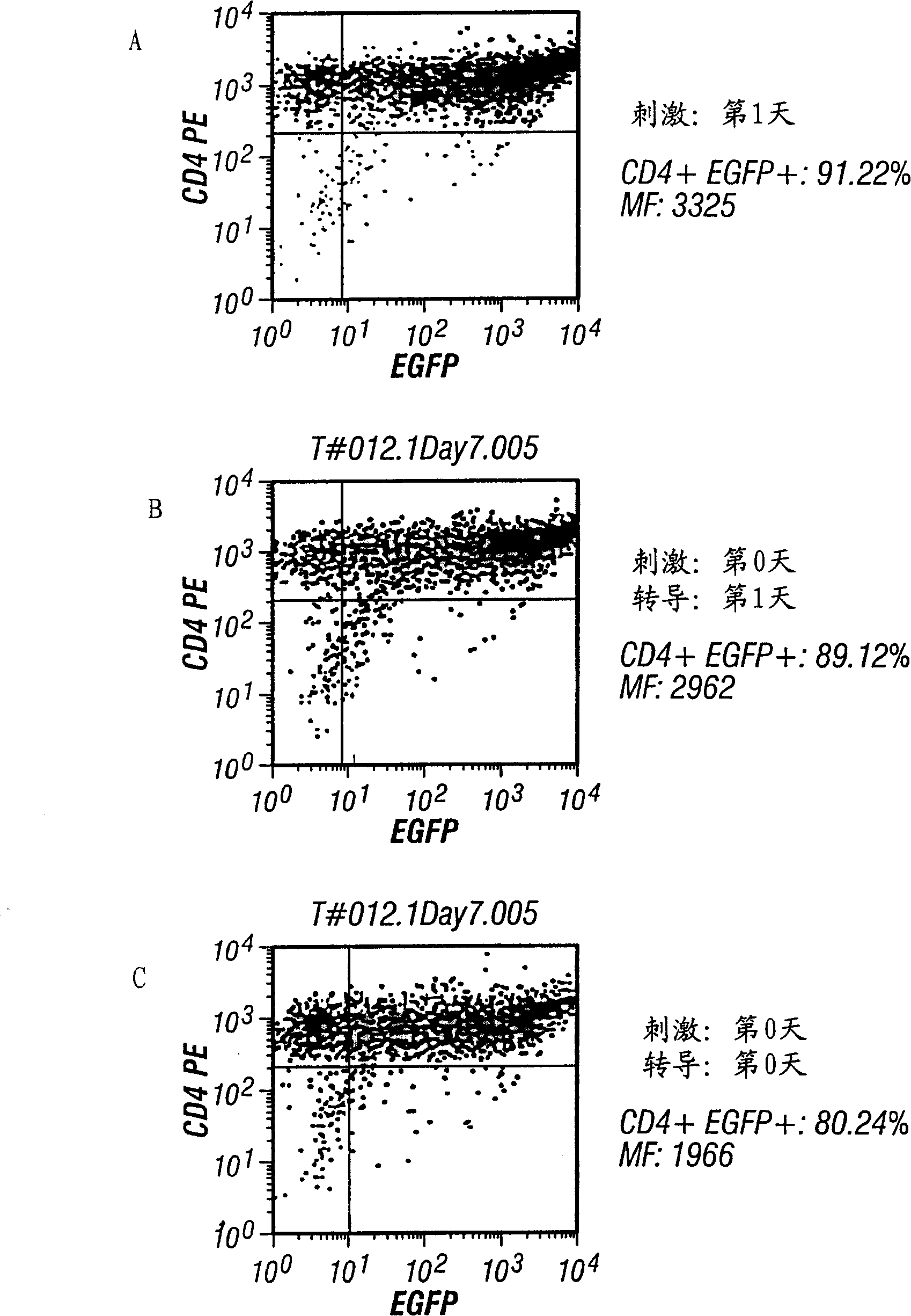
[0122] After transduction, and after 7 or 14 days in culture, cells were analyzed for CD4+ and / or green fluorescent protein (GFP) by flow cytometry.

[0123] A comparison of the above three transduction schemes can be seen figure 2 . After being transduced with pN2cGFP at MOI 20, it was contacted with αCD3 and αCD28 immobilized on microbeads, and the transduction rate was about 91%. The efficiency of bead contact followed by transduction was about 89%, and the transduction rate performed simultaneously was about 80%. In this experiment, CD4+ T cells were selected by monocyte adhesion removal method, CD14MACS removal method and CD4MACS enrichment method. Antibody immobilization was as follows. Contact with support at 37°C and 5% CO 2 under conditions. The culture condition is 500,000 CD4+T cells / ml, Yssel's medium containing 2% human serum albumin. FACS analysis was performed on day 7 after selection. MF indicates mean fluorescence. ...


PUM


Abstract

The present invention provides methods and compositions for efficiently transducing cells with viral vectors. Contacting the cells to be transduced with one or more cell surface binding molecules increases the rate of transduction. Contacting of the cells with the cell surface binding molecule can be performed before, after or simultaneously with the viral vector. Transduction vectors can be constructed to express a gene of interest, thereby rendering the transduced cells useful as therapeutic and prophylactic agents.

Description

technical field [0001] The present invention relates to methods and compositions for stably transducing cells with viral vectors. The method increases transduction efficiency by contacting the cells to be transduced with one or more cell surface binding molecules. The contacting step can be performed before, after or during the introduction of the viral vector into the cells. The invention also relates to the use of such stably transduced cells in other fields, including the expression of nucleic acids carried by vectors or the therapy of living organisms. Background technique [0002] "Transfection" generally refers to the technology of introducing genetic material into cells, which has made a significant contribution to the revolution of molecular and recombinant technology in the field of biology. Transfection techniques for higher eukaryotic cells include, for example, calcium phosphate precipitation, DEAE-dextran treatment, electroporation, microinjection, lipofection...

Claims


Application Information

Patent Timeline
Patent Type & Authority Patents(China)
IPC IPC(8): C12N15/87C12N15/86A61K48/00A61P31/12A61P31/18A61P35/00C12N15/09C12N1/15C12N1/19C12N1/21C12N5/00C12N5/08C12N5/10C12N7/00C12N15/63C12N15/867
CPCA61K48/00C12N15/86C12N2740/16043A61P31/12A61P31/18A61P35/00C12N15/87
Inventor L·休缪韩伟吕小宾V·斯德普西金M·莱舍B·戴维斯张永年B·德罗普利克
Owner VIRXSYS
Why Eureka
Try Eureka