Method and apparatus for examining hollow lumen by virtual endoscope

A technology of virtual endoscopy and inspection method, which is applied in the field of virtual endoscopy of the intestine, can solve the problems such as the lack of high recognition of the technology and too much redundant information.

Inactive Publication Date: 2007-11-14
SIEMENS AG
View PDF2 Cites 7 Cited by
  • Summary
  • Abstract
  • Description
  • Claims
  • Application Information

AI Technical Summary

Problems solved by technology

[0008] However, observers in virtual colonoscopies utilizing the manner in which this type of image data is displayed are burdened with excessive and often redundant information, rendering this technique less than highly acceptable in practice

Method used


Image

  • Method and apparatus for examining hollow lumen by virtual endoscope
  • Method and apparatus for examining hollow lumen by virtual endoscope
  • Method and apparatus for examining hollow lumen by virtual endoscope

Examples


Embodiment Construction

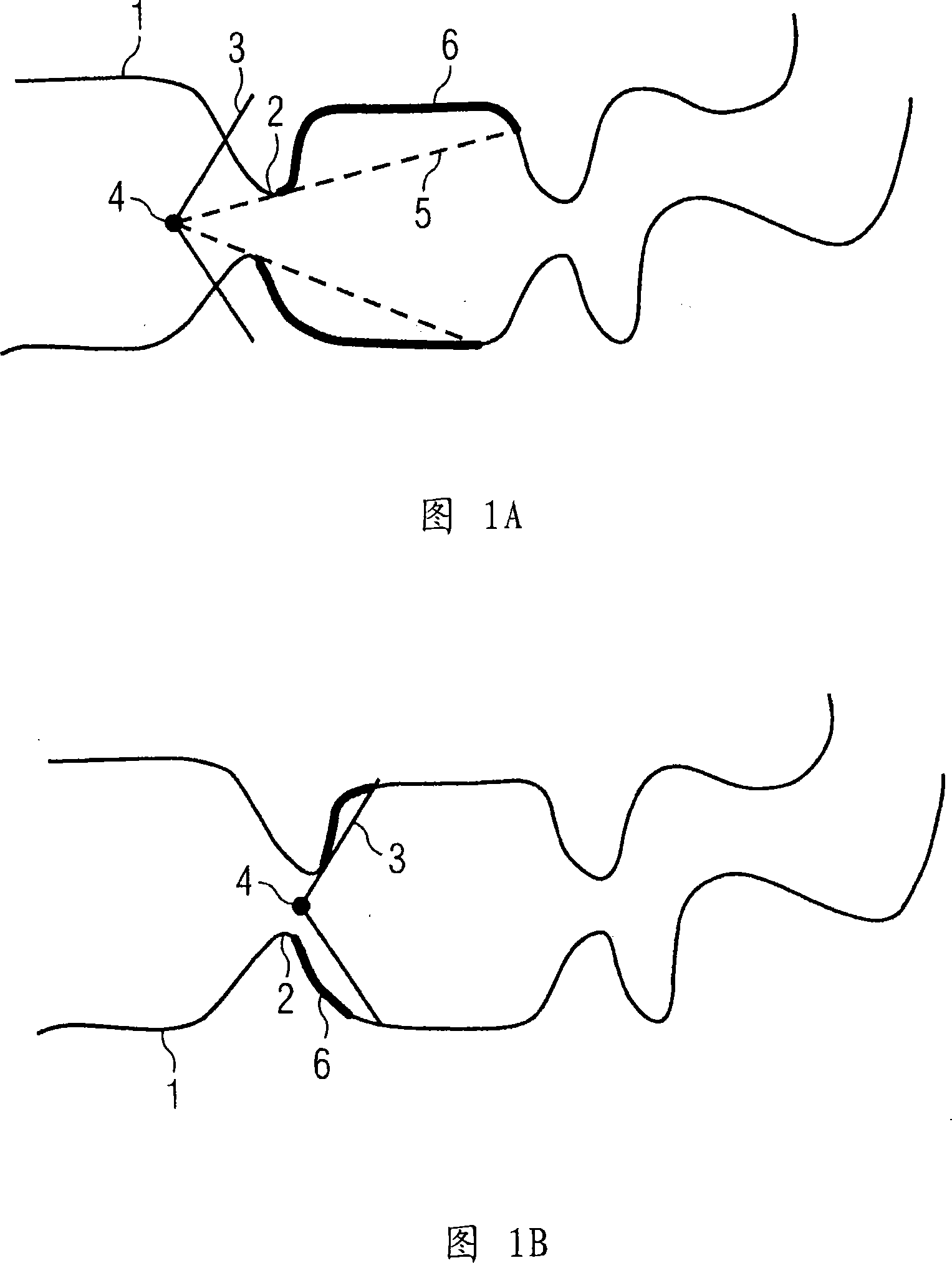
[0023] 1A and 1B show a limitation of the field of view during a virtual colonoscopy, which can be caused both by folds 2 in the intestinal wall 1 and by the predetermined field of view 3 of the virtual endoscope. The respective instantaneous positions 4 of the virtual endoscope with the field of view 3 of the endoscope or with the field of view 5 limited by the folds 2 are shown in the figures. In each case there are regions 6 which are invisible to the observer during this virtual traversal.

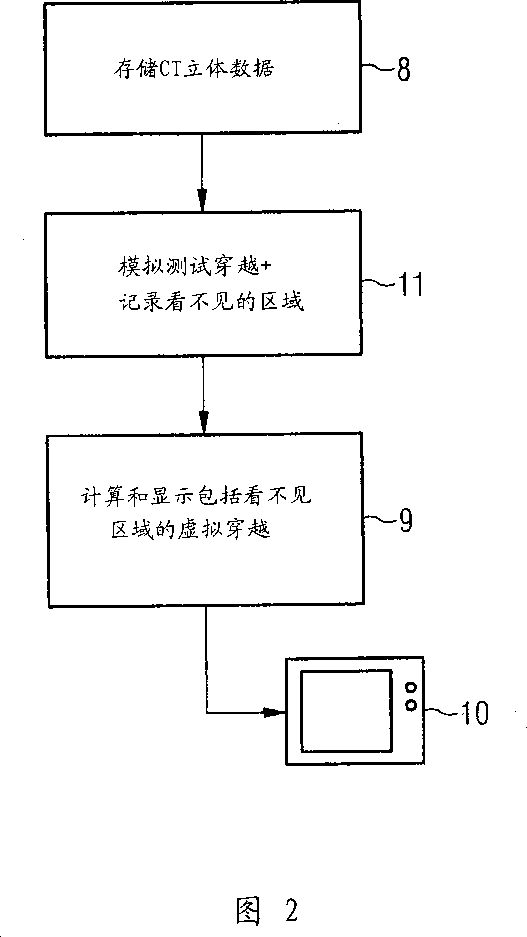
[0024] In the method according to the invention, a test crossing is initially simulated on the basis of volumetric data of the intestinal tract, which can originate, for example, from computed tomography recordings and are stored in the memory unit 8 of the device. As in every virtual traversal, here, in principle, the gut must be extracted from the volumetric data by suitable segmentation techniques. In addition, for later image representation or display, it is necessary to use volum...


PUM


Abstract

The invention relates to a method and a device for virtual endoscopy in a hollow tract, in which at least one volume record of the hollow tract (1), recorded by tomographic imaging, is provided from which a virtual flight through the hollow tract (1) in endoscopic perspective is calculated and visualized on a display device (10). In at least one embodiment, in the method and the device, before the visualization of the virtual flight, a virtual test flight without visualization is first simulated in which unobservable areas (6) of the hollow tract are detected during the test flight. Subsequently, the observer is notified of the unobservable areas (6) close to the location during the visualization of the virtual flight or the unobservable areas (6) are automatically visualized during the virtual flight. Using the present method and the associated device, in at least one embodiment, the time consumption during virtual endoscopy may be reduced for the observer without overloading him with redundant information.

Description

technical field [0001] The invention relates to a method and a device for virtual endoscopic examination of cavity organs, in particular of the intestinal tract, in which at least a three-dimensional data set of the cavity organs recorded by means of a tomographic imaging method is provided, from which A virtual traversal of the volume data set through the cavity organ is calculated in the endoscopic perspective or in a display derived therefrom and displayed on the display device. Background technique [0002] With imaging tomography methods, images of the interior of objects can be recorded and displayed in various ways. In this way, for example layer images or VRT images (VRT: Volume Rendering Technik) can be calculated and represented from the volume data sets obtained in this case. In addition, frequently used imaging tomography methods are, for example, computed tomography, magnetic resonance tomography or three-dimensional ultrasound imaging. [0003] In addition to...

Claims


Application Information

Patent Timeline
Patent Type & Authority Applications(China)
IPC IPC(8): A61B19/00G06Q50/00A61B6/03
CPCA61B19/50A61B2019/5289G06T2207/30028G06T19/00G06T2210/41G06T7/0012G06T2207/10081A61B34/10A61B2090/364G06T19/003
Inventor 卢茨·冈德尔
Owner SIEMENS AG
Why Eureka
Try Eureka