Quantitative Monte Carlo simulation method for light transfer characteristic in biological tissue

A technology of biological tissue and simulation method, which is applied in the application field of spectral technology, and can solve the problems of tissue model limitation, low precision, and inability to use biological tissue, etc.

Inactive Publication Date: 2008-10-15
HUAZHONG UNIV OF SCI & TECH
View PDF0 Cites 27 Cited by
  • Summary
  • Abstract
  • Description
  • Claims
  • Application Information

AI Technical Summary

Problems solved by technology

Among them, the 'MCML' method (S.L.Jacques and L.Wang. "Monte Carlo modeling of light transport intissues," in 1995 Proc OPTICAL-THERMAL RESPONSE OFLASER-IRRADIATED TISSUE Conf., PP.73-99. and L.Wang, S.L. Jacques, and L. Zheng, "MCML--Monte Carlo modeling of light transport inmulti-layered tissues, "Comput.Methods.Programs.Biomed., vol.47, pp.131-146, 1995.) is the most widely used in the world Broad and the most accurate method, but this method is only suitable for simplified biological tissues with col...

Method used


Image

  • Quantitative Monte Carlo simulation method for light transfer characteristic in biological tissue
  • Quantitative Monte Carlo simulation method for light transfer characteristic in biological tissue
  • Quantitative Monte Carlo simulation method for light transfer characteristic in biological tissue

Examples


Embodiment 1

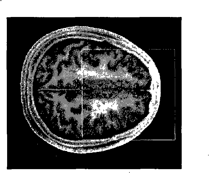
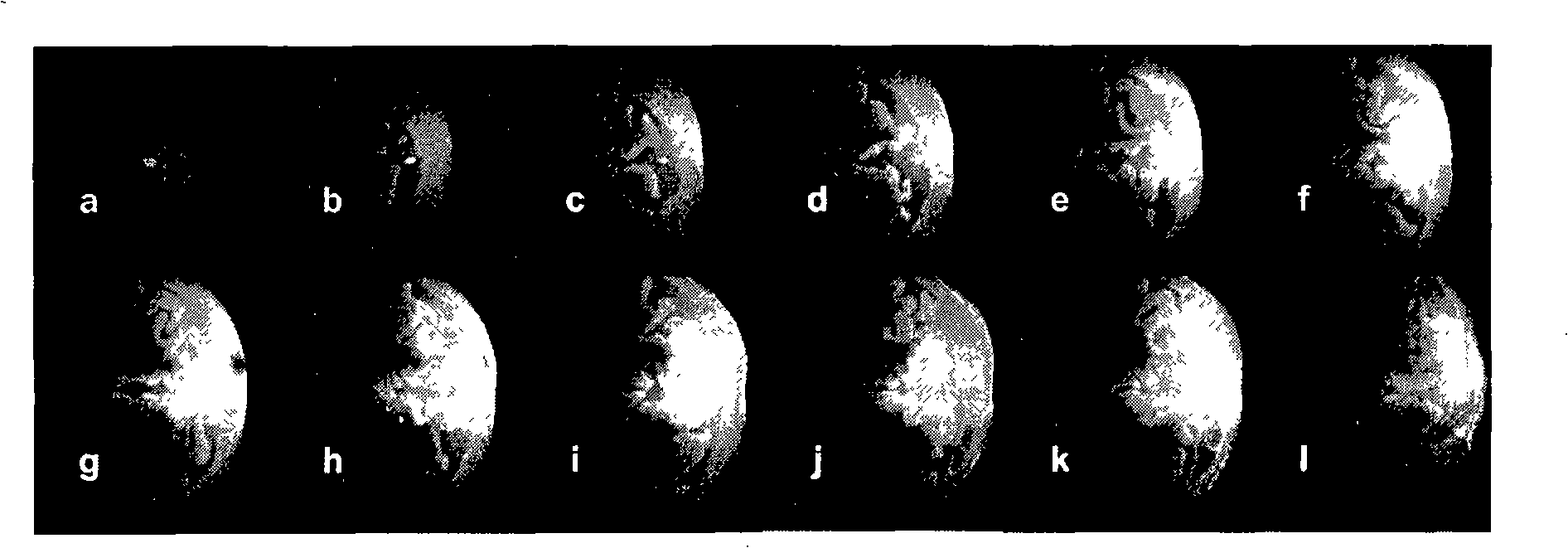
[0075] Take the voxel side length as 0.4mm and the number of photons as 10 7 And the tissue optical parameters in the following table use the method of the present invention to simulate light in such as figure 2 Transmission in the frontal brain of the digital human shown in the cross-sectional view, the resulting luminous flux distribution is as follows image 3 shown. image 3 The subgraph g in corresponds to figure 2 The area inside the white wireframe in . This tissue model cannot be simulated using the existing new method 'tMCimg' because the method cannot be applied to biological tissues with curved surfaces. The luminous flux distribution of any longitudinal section obtained by using the existing widely used 'MCML' method with the same parameters is as follows: Figure 4 shown. image 3 The distribution maps of different cross-sections are different, which is consistent with the three-dimensional tissue structure of brain tissue. Figure 4 The distribution of lu...

Embodiment 2

[0078] The accuracy of the method of the present invention is tested with a columnar multi-layer simplified tissue model, and compared with the results obtained by the existing 'MCML' method with the highest accuracy. The voxel side length is 0.5mm, and the number of photons is 10 5 . The optical parameters are shown in the table below.

[0079]

[0080]

[0081] The following table shows the comparison results of the diffusion coefficient of light transmitted in layered simplified biological tissues obtained by the method of the present invention and the existing method 'MCML' with the highest accuracy, and the new method 'tMCimg' which can be used in three-dimensional structural tissues. Figure 5The upper subgraph is the luminous flux distribution diagram, and the lower subgraph is the curve of light absorption changing with the depth of biological tissue. the table below and Figure 5 All show that the accuracy of the present invention is not lower than the level ...


PUM


Abstract

A quantitative Monte Carlo simulation method for transmission characteristics of light in a biological tissue pertains to the application of spectrum technology; the method firstly describes a spatial structure of the target biological tissue as a three-dimensional digital matrix, the surface characteristics of an incident light source are a set of photons with the set number, the position of the incident light source and the direction of the incident light are given to the initial position and the direction of each photon; the transmission process of each photon is tracked, and then a light absorption matrix and the information of all escaped photons are output. The method quantitatively simulates the light transmission characteristics in any biological tissue with three-dimensional structure by establishing a Monte Carlo model of transmitting the light in the tissue. The method has high precision and good stability, which can obtain the rich characteristics of the light transmission in the real biological tissue. The application of the method can optimize the design of a biological tissue optical detection/imaging system, provide precise information for the dose quantification for light treatment and provide an accurate and reliable tool for the positioning of a detection area and the quantitative analysis of signals in the biological tissue by using the spectrum technology.

Description

technical field [0001] The invention belongs to the fields of spectral technology application, mathematical simulation and biomedical engineering, and relates to a quantitative Monte Carlo simulation method of light transmission characteristics in tissues. Background technique [0002] The application of spectroscopic technology to the detection and imaging of biological tissues has the advantages of non-invasive measurement, high time resolution, and low cost compared to existing mature technologies such as PET, fMRI, EMG / ECG / EEG, etc. It is of great significance to disease diagnosis and monitoring, optical clinical treatment, brain function mechanism and physiological and pathological research. In recent years, spectroscopic technology has been widely praised in the research and clinical application of biological tissue detection imaging, but its accuracy and reliability have not been widely accepted. The reason is that light, especially the most commonly used near-infrare...

Claims


Application Information

Patent Timeline
IPC IPC(8): G06F19/00A61B5/00A61N5/06
Inventor 骆清铭龚辉李婷
Owner HUAZHONG UNIV OF SCI & TECH
Why Eureka
Try Eureka