Human papilloma virus (HPV) detection using nucleic acid probes, microbeads and fluorescent-activated cell sorter (FACS)

A probe, nucleic acid capture technology, used in the determination/inspection of microorganisms, particle and sedimentation analysis, measuring devices, etc., can solve the problems of indistinguishable, unsatisfactory cervical cancer risk, etc.

Inactive Publication Date: 2009-01-07
GENERA BIOSYSTEMS LTD
View PDF0 Cites 12 Cited by
  • Summary
  • Abstract
  • Description
  • Claims
  • Application Information

AI Technical Summary

Problems solved by technology

However, the Pap smear is a somewhat unsatisfactory assay for definitively determining cervical cancer risk because the technique h

Method used


Image

  • Human papilloma virus (HPV) detection using nucleic acid probes, microbeads and fluorescent-activated cell sorter (FACS)
  • Human papilloma virus (HPV) detection using nucleic acid probes, microbeads and fluorescent-activated cell sorter (FACS)
  • Human papilloma virus (HPV) detection using nucleic acid probes, microbeads and fluorescent-activated cell sorter (FACS)

Examples


Embodiment 1

[0274] Example 1: HPV Diagnosis - DNA Isolation and Amplification

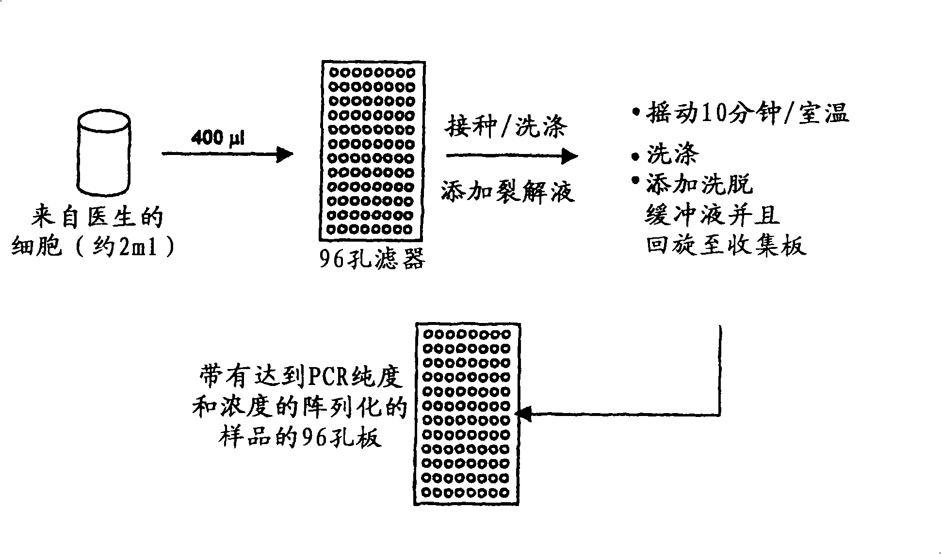
[0275] exist figure 2 An overview of DNA extraction methods used to isolate DNA in HPV diagnostic methods is shown in .

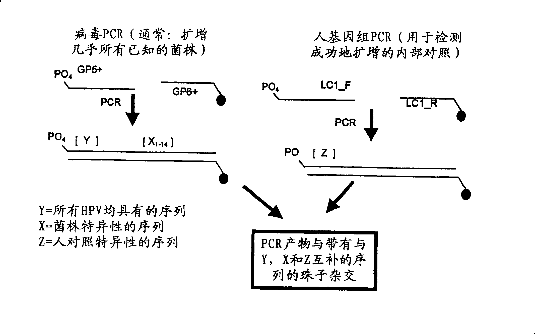
[0276] Such as image 3 DNA samples were amplified using PCR as indicated. Primers GP5+ and GP6+ were used to generate amplicons of any HPV strain present in the DNA sample. Primer GP6+ contains a fluorescent label, more specifically Cy5, to allow for later visualization of the binding of the amplicon to the binding reagent. The resulting viral amplicon contains a conserved region (Y), which is conserved among all HPV strains examined, and a region X that is variable (i.e., strain-specific) between HPV strains n , where n represents the variable region associated with each HPV strain. The binding partners immobilized on the beads will specifically bind the HPV strain-specific genome.

[0277] In addition, amplicons from human subject genomic DNA were also generated using LC1_F and LC...

Embodiment 2

[0278] Example 2: HPV Diagnosis - Multiplex Detection

[0279] The amplicons generated in Example 1 were hybridized to an array of binding reagents each carrying a protein from each HPV strain c, 11, 16, 18, 31, 33, 35, 42, 45, 51, 52 , 56, 58, 59, 67 and 68 (X 1 to X 16 ) produced a polynucleotide complementary to the variable region of a putative viral amplicon. For the nucleotide sequence of the capture nucleic acid immobilized on the beads see Figure 9 . Furthermore, the array comprises binding reagents comprising polynucleotides complementary to the conserved region (Y) of the HPV viral amplicon. Finally, a binding reagent comprising a polynucleotide complementary to the human control amplicon sequence is included. Capture nucleic acid can be DNA or RNA. If RNA is used, reverse transcriptase may be required to generate RNA from DNA amplicons.

[0280] Each binding reagent in the array comprises microspheres or beads with different sizes and different fluorescent (T...

Embodiment 3

[0284] Example 3: Comparison of multiple detection methods and traditional HPV diagnosis

[0285] Table 5 below provides an overview comparing the multiplex HPV detection method of the present invention with existing histological methods for HPV diagnosis.

[0286] Table 5 Comparison of HPV diagnostic methods

[0287] HPV diagnostic methods


PUM


Abstract

The present invention relates generally to the field of diagnostic and detection assays. More particularly, the present invention provides methods, and reagents including biochips for detecting the presence of, or distinguishing between, one or more analytes in a sample.

Description

field of invention [0001] The present invention relates generally to the field of diagnostic and detection assays. More specifically, the present invention provides methods and reagents, including biochips, for detecting the presence or distinguishing one or more analytes in a sample. Background technique [0002] At the end of the specification, bibliographic details for references provided in this application are listed. [0003] Any prior art in this specification is not and should not be considered as an acknowledgment or any form of suggestion that such prior art forms part of the common general knowledge in any country. [0004] The need for screening methods that rapidly and reliably detect multiple analytes in a single assay is critical not only in the field of clinical diagnostics, but also in screening applications such as screening for environmental toxins, and drug screening. [0005] One such area where improved screening methods and reagents are urgently need...

Claims


Application Information

Patent Timeline
IPC IPC(8): C12Q1/68G01N15/14C07H21/02G01N33/48C12P19/34
Inventor K·波特T·戈尔德
Owner GENERA BIOSYSTEMS LTD
Why Eureka
Try Eureka