Method and system for guiding operation of electronic endoscope by auxiliary computer

An electronic endoscope and computer-aided technology, applied in the fields of radiological diagnosis instruments, medical science, surgery, etc., can solve problems such as tissue or position misjudgment, and achieve the effect of avoiding repeated viewing.

Inactive Publication Date: 2009-03-04
SHENZHEN GRADUATE SCHOOL TSINGHUA UNIV
View PDF0 Cites 49 Cited by
  • Summary
  • Abstract
  • Description
  • Claims
  • Application Information

AI Technical Summary

Problems solved by technology

[0003] Although the current medical electronic endoscope technology has been widely used, but currently using medical electronic endoscope technology in clinical practice, clinicians can only see the partial two-dimensional images captured by the endoscopic probe. The locatio...

Method used


Image

  • Method and system for guiding operation of electronic endoscope by auxiliary computer
  • Method and system for guiding operation of electronic endoscope by auxiliary computer
  • Method and system for guiding operation of electronic endoscope by auxiliary computer

Examples


Embodiment Construction

[0019] The present invention will be described in detail below according to the drawings and embodiments.

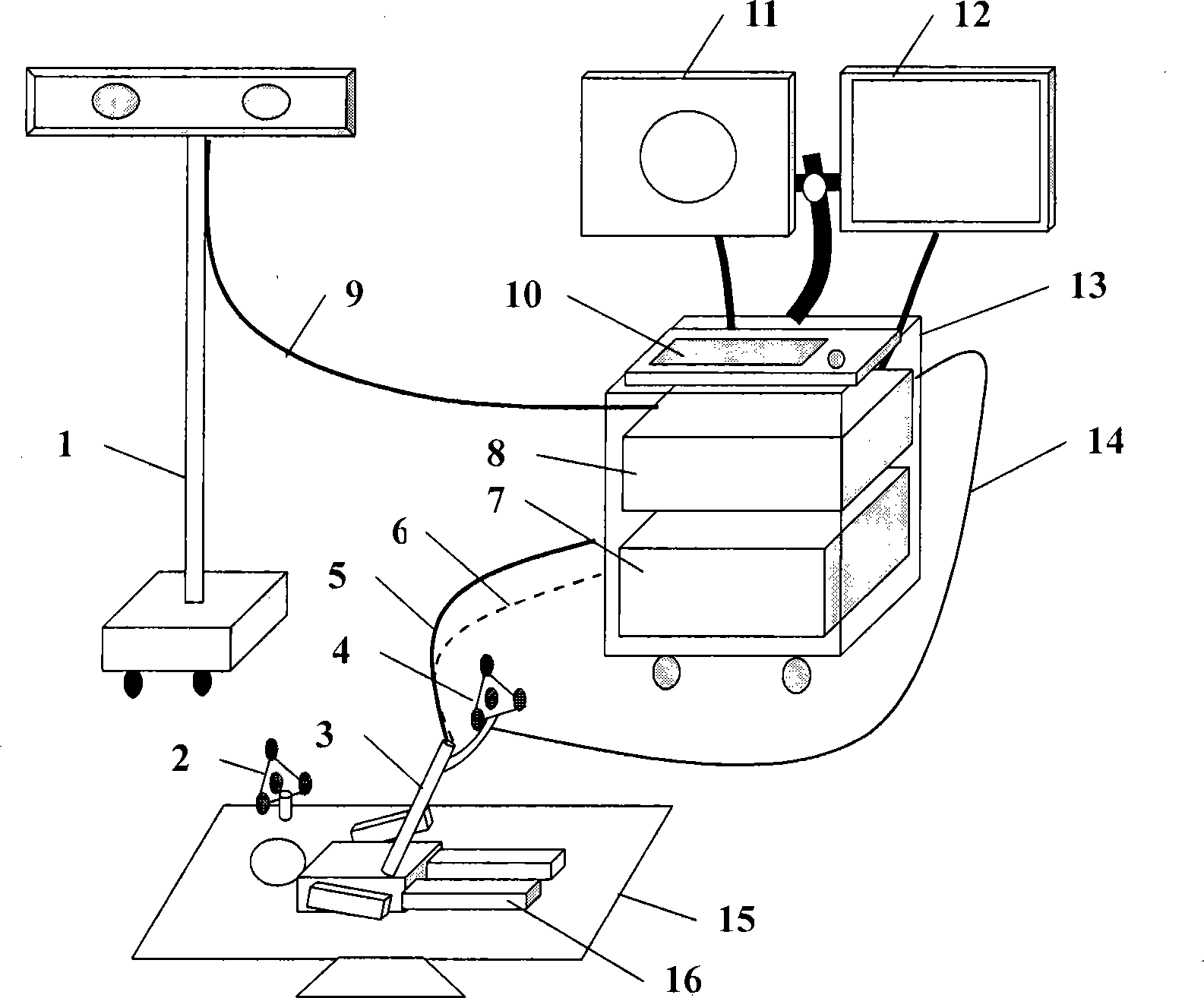
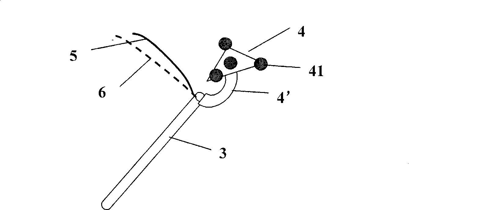
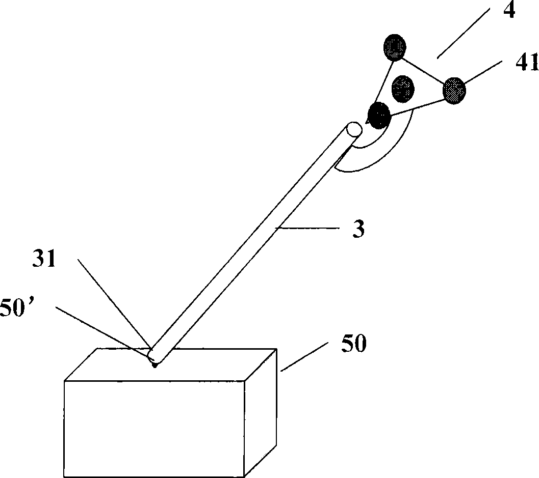
[0020] refer to figure 1 , the computer-aided system for guiding electronic endoscope operation mainly includes: an electronic endoscope system for photographing and displaying the image of the position where the endoscopic probe arrives, for tracking the position and direction of the endoscopic probe for spatial positioning system, image workstation and supporting image navigation processing software. Wherein, the electronic endoscope system includes an endoscope host 7, an endoscope probe 3 and an endoscope display 11, and the endoscope probe 3 is connected with the endoscope host 7 through an optical fiber 5 for connecting the endoscope probe 3 The environment image around the top is transmitted to the endoscope host 7, and the endoscope probe 3 is connected to the endoscope host 7 through the optical cable 6, so that the light source illuminates the tissue around th...


PUM


Abstract

The invention discloses a method and a system for introducing an electronic endoscope into operation with the assistance of a computer. The system comprises an electronic endoscope system, a space orientation system used for tracking a position and a direction of the probe head of the endoscope, an image workstation used for producing a planar or three-dimensional image of a surgery area of a patient according to an input image and blending and displaying an image of the probe head of the endoscope and the planar or three-dimensional image by connecting with the space orientation system, and auxiliary image orientation and processing software. The system tracks the position and the direction of the probe head of the endoscope through a space track technology and a computerized image processing technology, so that by combing the planar image with the image of the endoscope with the assistance of images as CT or X-ray pictures, a doctor can not only see the planar image captured by the probe head of the endoscope, but also see the position of the probe head of the endoscope and the conditions of the peripheral tissue. Therefore, the doctor can fully control the comparative position of the probe head of the endoscope in the body, thereby improving the quality of endoscope surgery and shortening the time of a surgery.

Description

technical field [0001] The invention relates to medical operation navigation equipment, in particular to the problem of using medical two-dimensional or three-dimensional images to complete the positioning of the apex of the medical endoscope probe in the body. Background technique [0002] As minimally invasive surgical techniques are increasingly adopted by doctors, endoscopic surgery has developed rapidly. At present, there are three types of endoscopic examinations and operations in hospitals: the first is to simply use an endoscope, and its disadvantages are that it can only be seen by the operator, the image is small, and the operation is not well coordinated, which is not conducive to teaching; The mirror is connected to the TV through a CCD camera, and its disadvantages are that the image is not clear, it cannot store and manage medical records, and it cannot print graphic reports; the third type is medical electronic endoscope. At present, the medical electronic en...

Claims


Application Information

Patent Timeline
IPC IPC(8): A61B17/00A61B1/00A61B6/00
Inventor 吴剑黄毅斌叶大田
Owner SHENZHEN GRADUATE SCHOOL TSINGHUA UNIV
Why Eureka
Try Eureka