Method and equipment for obtaining full-field ultrasound scan image data

An image data, full field of view technology, applied in the field of medical imaging technology products, can solve problems such as hindering the development of standardization and standardization of ultrasound examination, scanning images can only display local measured target tissues at the same time, and large image differences.

Inactive Publication Date: 2009-07-08
UNITED IMAGING SYST BEIJING CO LTD
View PDF0 Cites 15 Cited by
  • Summary
  • Abstract
  • Description
  • Claims
  • Application Information

AI Technical Summary

Problems solved by technology

[0003] Hand-held inspection is firstly affected by the size of the probe. The imaging range of a single scan is very small. The scanning width of the probes equipped today is generally not greater than 80mm, and the volume probe is not greater than 40mm*80mm, which limits the field of view and makes the scanned image only at the same time. Display the local target tissue under test; secondly, it is affected by the technique and experience of the examiner, especially when scanning a large part or multiple parts, it needs to move multiple times, and the examinations are performed by different examiners and at different times It is not easy to achieve consistency; the re-scanning process cannot be completed automatically, and the positional relationship of the acquired multi-frame images is uncertain, and it is impossible to form a panoramic image through reconstruction; the final scanning process is relatively long, and the examiner must perform a hand-held scan while making a diagnosis
[0004] The above factors make it impossible for traditional ultrasound examination to obtain panoramic images of the measured area, and the images obtained by the inspectors are quite different. The comparison of X-ray, CT, MRI, anatomical images, etc. hinders the standardization and normalization of ultrasound examination, and is not suitable for ultrasound screening of large-scale population

Method used


Image

  • Method and equipment for obtaining full-field ultrasound scan image data
  • Method and equipment for obtaining full-field ultrasound scan image data
  • Method and equipment for obtaining full-field ultrasound scan image data

Examples


Embodiment Construction

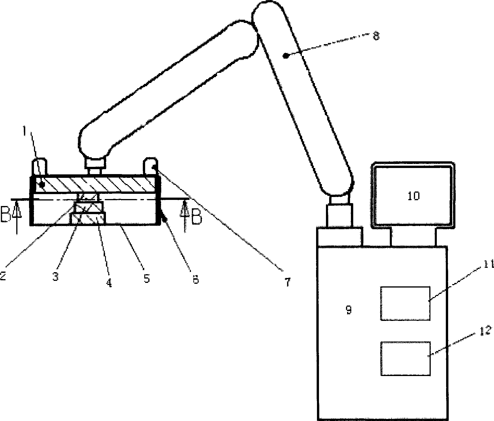
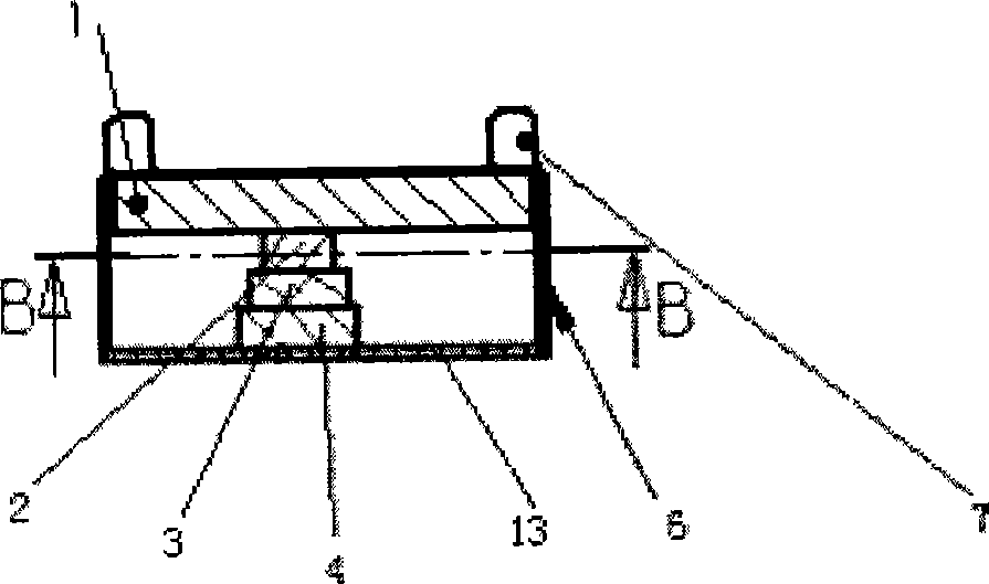
[0026] figure 1 is a structural schematic diagram of the device of the present invention, figure 2 yes figure 1 Sectional view along line B-B. The equipment of the present invention comprises: a scanning bed, which has a body position fixing device of the measured object, and the body position fixing device can move back and forth or left and right along the scanning bed to adjust the position; a scanning platform 6 is a rectangular frame with a frame, the scanning The inspection platform 6 can be made of strong plastic material, and the plane of the scanning platform 6 in contact with the target tissue to be measured is the scanning surface 5 . During scanning, the scanning surface 5 covers the target tissue to be scanned. The upper part of the scanning platform 6 is fixedly connected to one end of an adjustable rocker arm frame 8 . The rocker arm frame 8 is connected to an ultrasound scanning control console 9 , on which there is a display 10 , and a central processing ...


PUM


Abstract

The invention relates to a method for obtaining the data of a total field of view ultrasound scanning image and a device thereof. The device comprises a large scale scanning platform which can cover the objective organization of a whole tested object. The scanning platform is internally provided with a scanning probe which is controlled by a central processing unit and obtains the data of the image by scanning.

Description

technical field [0001] The invention belongs to the field of medical imaging technology products, and in particular relates to an ultrasonic inspection device and a related method, which can obtain ultrasonic inspection of full-field ultrasonic scanning image data. Background technique [0002] Traditional ultrasound examinations all use hand-held methods. The examiner holds the probe, places the probe on the surface of the target tissue to be measured, and scans the target tissue by moving the probe position or changing the scanning angle to obtain irregular dynamic two-dimensional images from different angles. dimensional image. [0003] Hand-held inspection is firstly affected by the size of the probe. The imaging range of a single scan is very small. The scanning width of the probes equipped today is generally not greater than 80mm, and the volume probe is not greater than 40mm*80mm, which limits the field of view and makes the scanned image only at the same time. Displ...

Claims


Application Information

Patent Timeline
Patent Type & Authority Applications(China)
IPC IPC(8): A61B8/00
Inventor 牟晓勇叶迪晏书君
Owner UNITED IMAGING SYST BEIJING CO LTD
Why Eureka
Try Eureka