

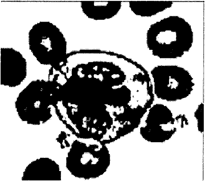
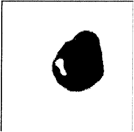
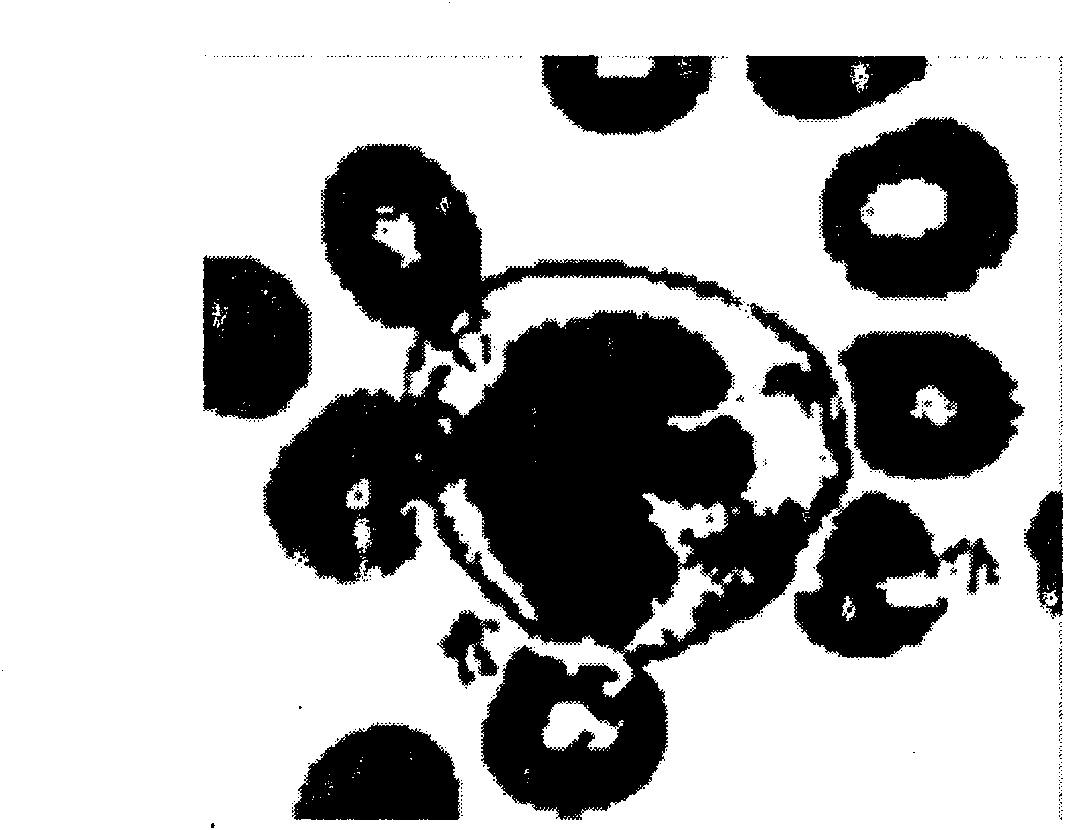
Segmentation method of cell nucleolus and cell membrane based on mixed contour model
A contour model, cell nucleolus technology, applied in the field of biological cell microscopic image segmentation, can solve the problems of poor segmentation effect and low precision
- Summary
- Abstract
- Description
- Claims
- Application Information
AI Technical Summary
Problems solved by technology
Method used
Image

Examples
Embodiment Construction
[0039] The present invention will be further described below in conjunction with the accompanying drawings.
[0040] A method for segmenting cell nucleoli and cell membranes based on a mixed contour model, the segment method comprising the following steps:
[0041] 1), the cell image to be segmented according to the mixed contour model to establish an energy function, the form is as follows:
[0042] E. E (φ, c 1 , c 2 , f 1 , f 2 ) = E M (φ, c 1 , c 2 , f 1 , f 2 )+P(φ)+L(φ) (11);
[0043] Among them, E M (φ, c 1 , c 2 , f 1 , f 2 ) represents the energy function based on the regional model and the local binary fitting model, and its calculation formula is:
[0044] E. M (φ, c 1 , c 2 , f 1 , f 2)=(1-λ g )E LBF (φ, f 1 , f 2 )
[0045] +λ g (x, y)E G (φ, c 1 , c 2 ) (7);
[0046] where λ g is the strategy weight parameter, E LBF (φ, f 1 , f 2 ) as defined in formula (6), E G (φ, c 1 , c 2 ) as defined in formula (5):
[0047] E. G (φ, c...
PUM
 Abstract
Description
Claims
Application Information