
Method for displaying soft copy of mammary gland X-line graph

A display method and line image technology, applied in image enhancement, image data processing, instruments, etc., can solve problems such as different, inability to clearly express diagnostic information, and reduce visible information

Inactive Publication Date: 2010-03-10
NINGBO UNIV
View PDF0 Cites 10 Cited by
  • Summary
  • Abstract
  • Description
  • Claims
  • Application Information

AI Technical Summary

Problems solved by technology

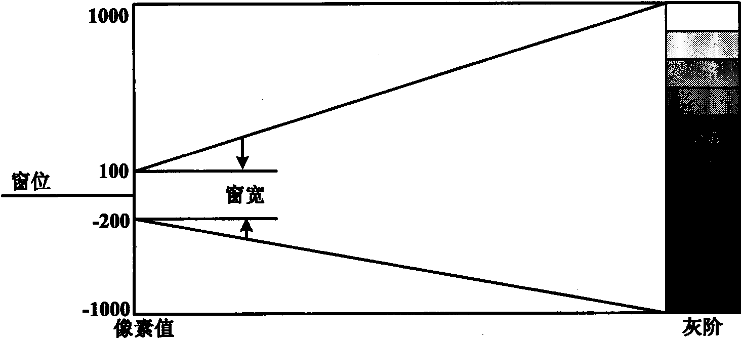
The window width and window level technology essentially achieves a better display effect by narrowing the visible pixel value range, but this method greatly reduces the visible information, the most obvious is that the mammogram cannot be clearly displayed at the same time. Outline and breast parenchyma area, in order to solve this problem, it is necessary to perform window adjustment operation, that is, to continuously observe mammogram images under different window width and window level parameters
At present, most window width and window level methods use a linear mapping method from the pixel value area to the displayable gray scale. However, different pixel values ​​represent different parts of the breast tissue, and the importance of the diagnostic information they contain is also different. Some window width and window level methods cannot reflect the different importance of different pixel values ​​in diagnosis, so they cannot achieve clear expression of diagnostic information

Method used


Image

  • Method for displaying soft copy of mammary gland X-line graph
  • Method for displaying soft copy of mammary gland X-line graph
  • Method for displaying soft copy of mammary gland X-line graph

Examples


Embodiment Construction

[0041] The present invention will be further described in detail below in conjunction with the accompanying drawings and embodiments.

[0042] Mammography images need to be converted from pixel values ​​to displayable gray scales to generate displayable images. The conversion process from pixel values ​​to displayable gray scales is as follows: figure 1 shown. When the mammogram image is initially displayed, the usual practice is to linearly map the selected pixel value area to the visible gray range of 0-255, so no matter what information the pixel value expresses in the image, the pixel value area of ​​equal length Maps to the same number of visible grayscales. The basic theory of the present invention is to allocate visible gray levels according to the importance of diagnostic information contained in different pixel value areas, thereby improving image display effect. A soft copy display method of a mammogram of the present invention comprises the following specific step...


PUM


Abstract

The invention discloses a method for displaying soft copy of a mammary gland X-line graph, in which a weighting function used for representing the importance of pixel values in a mammary gland zone and a monotonic increasing dynamic range conversion function are constructed by performing statistics to obtain a probability density column diagram of pixel value x in the mammary gland zone of the mammary gland X-line graph, each pixel value in the mammary gland X-line graph is mapped to displayable grayscale by using the dynamic range conversion function to form a display graph, substantially each pixel value in the mammary gland X-line graph is subject to non-linear mapping to displayable grayscale, as the mapping relationship adopts non-linear mapping, the result obtained by the invention can show different importance of message of different mammary gland tissue parts, thus realizing clear expression of important diagnostic message; in addition, by display graph by the method of the invention, the display effect of the mammary gland substantial zone can be vastly improved, and simultaneously the display effect of the mammary gland outline can be maintained, thus keeping distinct andvisible mammary gland outline.

Description

technical field [0001] The invention relates to a display method of digital medical images, in particular to a soft copy display method of mammogram images. Background technique [0002] The display effect of images is very important for diagnosis. Mammograms often contain fuzzy diagnostic information on a small scale. The use of advanced soft copy display technology can improve the conspicuousness of case features in mammograms. Mammography images generally follow the medical digital image communication standard (DICOM3.0), which was jointly formulated by the North American College of Radiology (ACR) and the Electrical Manufacturers Association (NEMA) in 1993, and is a de facto industry standard. The pixel values ​​of mammography images vary widely, and generally each pixel value is stored in 10-16 bits, but the input range provided by the display device is fixed, and the general display has only 256 gray levels, so DICOM (Digital Imaging and Communication) is required. of...

Claims


Application Information

Patent Timeline
IPC IPC(8): A61B6/03G06T5/00
Inventor 李均利魏平陈刚金林鹏祝卫峰裘意娜汪永生
Owner NINGBO UNIV
Learn More