Method for enhancing medical X-ray image display effect

An image display and X-ray technology, applied in the field of digital images, can solve problems such as blurred images and inability to reflect image detail information, and achieve the effect of enhancing contrast, improving image tissue edge contrast, and improving image detail signals

Inactive Publication Date: 2010-07-21
XIAN HWATECH MEDICAL INFORMATION TECH
View PDF0 Cites 23 Cited by
  • Summary
  • Abstract
  • Description
  • Claims
  • Application Information

AI Technical Summary

Problems solved by technology

This method better solves the problem of image blur and provides a good idea for image en

Method used


Image

  • Method for enhancing medical X-ray image display effect
  • Method for enhancing medical X-ray image display effect
  • Method for enhancing medical X-ray image display effect

Examples


Embodiment Construction

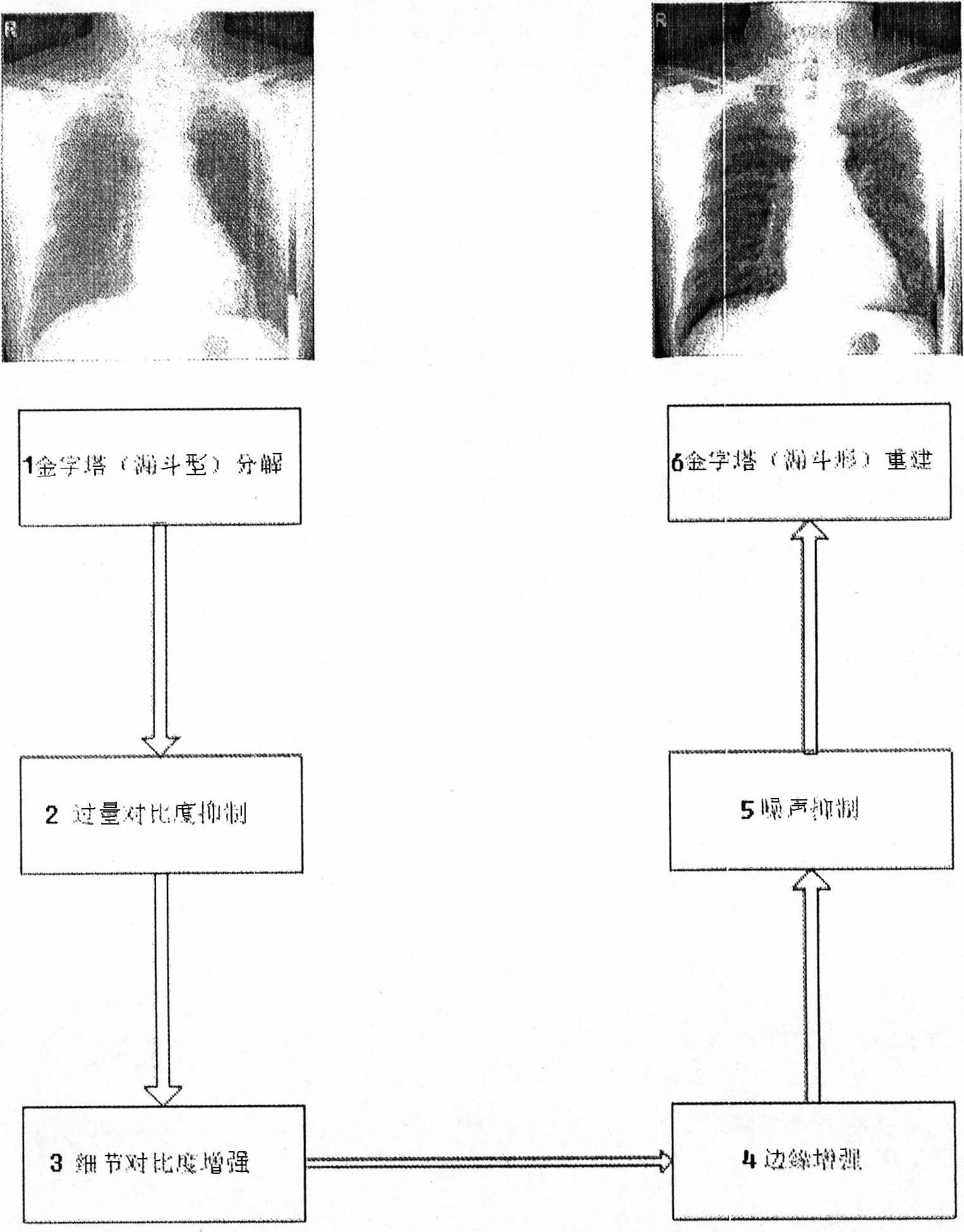
[0032] An embodiment is narrated below in conjunction with accompanying drawing, and the present invention is further described.

[0033] This embodiment is accomplished through the following method steps: image pyramid (funnel-shaped) decomposition, excessive contrast suppression, detail contrast enhancement, edge enhancement, noise suppression, pyramid (bucket-shaped reconstruction), such as figure 1 shown.

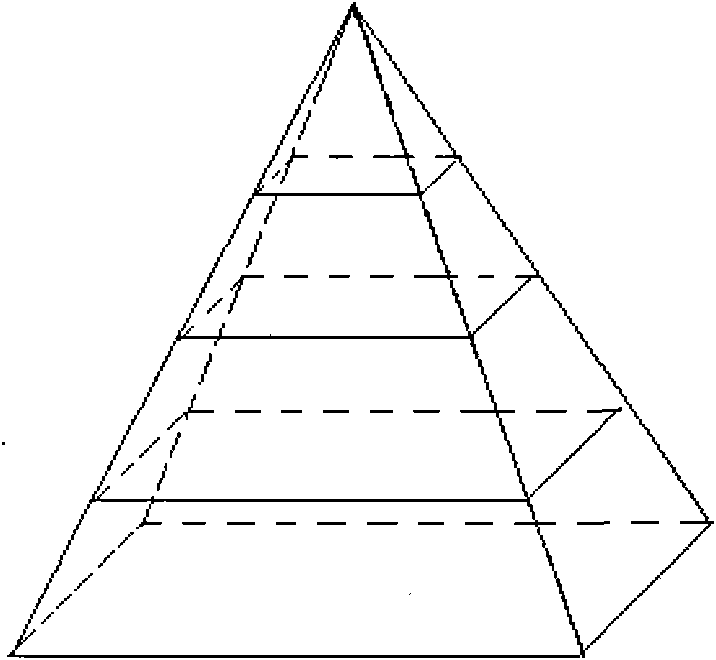
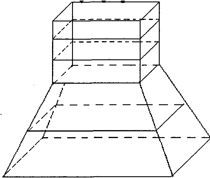
[0034] .1 Image pyramid (funnel-shaped) decomposition:

[0035] Using the Laplacian pyramid decomposition transformation, the medical image is decomposed into a pyramid-like bucket arrangement, and the 4096*4096 image is decomposed into 12 layers of resolution images (2 12 =4096), if the image is not square, take the maximum length and width. The image is interlaced and alternated, and the next-level low-resolution image is obtained by sampling. And so on (i=0....L-1). Such as diagram 2-1 However, the difference from pyramid decomposition is that when the image is...


PUM


Abstract

The invention discloses a method for enhancing medical X-ray image display effect, and relates to a digital image technology. The image enhancing method is completed through the following methods and steps: image pyramid (funnel-shaped) decomposition, excessive contrast inhibition, detail contrast enhancement, edge enhancement, noise suppression and pyramid (funnel-type reconstruction). By adjusting the pixel dynamic range distribution of an image, the method improves the detail signal of the image, enhance the contrast, the clarity and the permeability of the digital image, suppress the noise, and realize the effect of image organization edge contrast improvement, organization detail performance and permeable and visible overall image organization. The method for enhancing medical X-ray image display effect is particularly applicable to computer-controlled X-ray CT scanners or medical radiation image processing systems.

Description

Technical field: [0001] The invention relates to digital image technology, and is especially suitable for computer-controlled X-ray tomographic scanning device or medical radiation image processing system. Background technique: [0002] For some image systems, unlike other signal systems - it is necessary to process or control the signal itself, but the observer has to make an evaluation based on the provided image. For example, in medical images, at present, the cause of the lesion cannot be obtained through the self-processing of the image itself, and the image must finally be observed and evaluated by the doctor to make a judgment. Therefore, whether the quality of the final image is good or bad greatly affects the correct judgment of the doctor on the lesion. Obviously, the image with poor quality is likely to cause the doctor to misjudge or miss a judgment, which is obviously not good for the diagnosis and treatment of the disease. of. [0003] X-ray images are formed...

Claims


Application Information

Patent Timeline
IPC IPC(8): A61B6/02G06T5/00
Inventor 程亮白霖抒马天德侯云涛王洪涛
Owner XIAN HWATECH MEDICAL INFORMATION TECH
Why Eureka
Try Eureka