Special clamping device for zygomatic fracture reduction surgery

A clamping device, a technique for zygomatic fractures, applied in the directions of surgery, medical science, etc., can solve the problems of secondary damage to the facial appearance, difficulty in fixing the zygomatic bone, unable to reset the zygomatic bone, etc., and achieves increased flexibility, a simple and reasonable structure, The effect of convenient operation

Inactive Publication Date: 2011-12-28
温州医科大学附属口腔医院
View PDF0 Cites 0 Cited by
  • Summary
  • Abstract
  • Description
  • Claims
  • Application Information

AI Technical Summary

Problems solved by technology

But at the part where the zygomatic bone is adjacent to the maxillary bone, because the bone wall of the maxillary sinus is very thin, it is difficult for the sunken zygomatic bone to reset the maxillary process of the zygomatic bone with general force; at the part where the zygomatic bone is adjacent to the frontal bone, because There is an eyeball in the orbital wall, and the inner side of the orbital wall is smooth, so there is no place to apply prying force to reset the displaced zygomatic frontal process; as for the displacement of the zygomatic process, the general lifting method can only reset the zygomatic arch The shape of the cheekbone cannot be completely reset as a whole, which often brings great difficulties to the final fixation of the cheekbone due to inaccurate reduction.
In addition, using a retractor or a crowbar, multiple incisions must be made on the skin tissue around the cheekbones, which in turn causes unnecessary secondary damage to the facial appearance of the injured

Method used


Image

  • Special clamping device for zygomatic fracture reduction surgery
  • Special clamping device for zygomatic fracture reduction surgery
  • Special clamping device for zygomatic fracture reduction surgery

Examples


Embodiment Construction

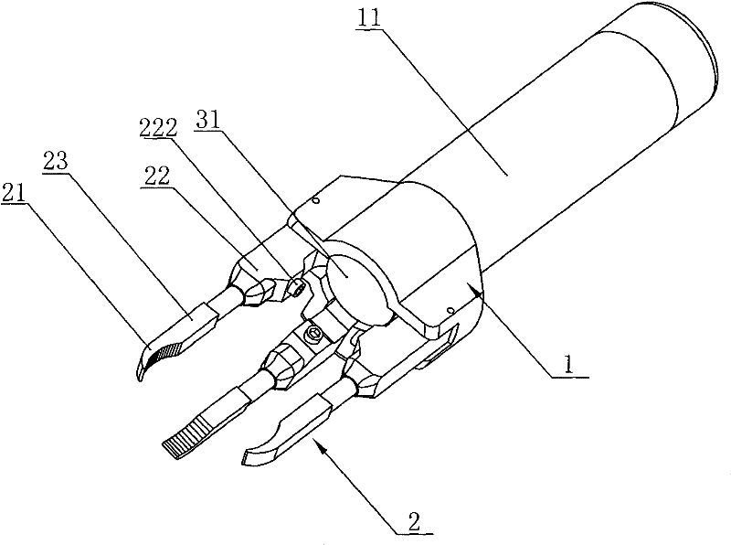
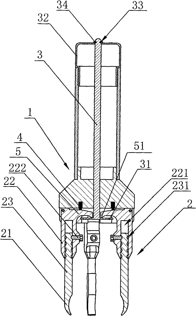
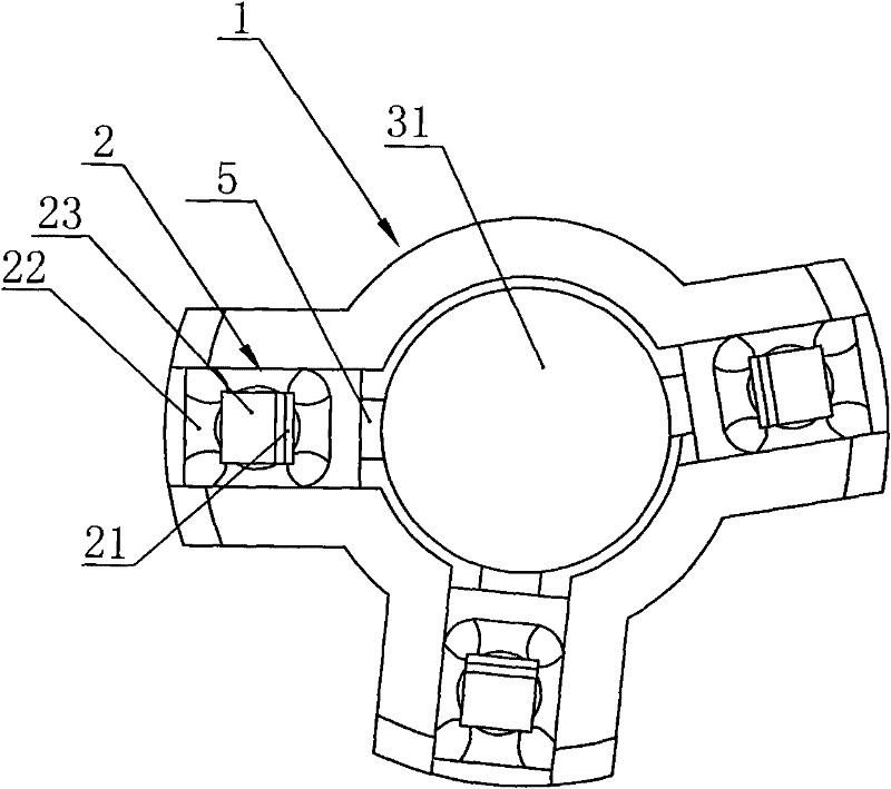
[0028] Such as Figure 1-Figure 5 As shown, the embodiment of the present invention is a special clamping device for zygomatic fracture reduction surgery, which includes a fixing base 1 and three clamping bodies 2, the fixing base 1 is fixed with a handle 11, and the clamping bodies 2 are hinged on the fixed On the seat 1, the clamping end of the clamping body 2 is provided with a clamping head 21 for realizing the clamping action, and the three clamping heads 21 are respectively connected to the inner surface of the outer lower edge of the orbit 6, the corner 7 of the outer edge of the orbit and the cheekbone, The corner 8 between the zygomatic bone and the zygomatic alveolar pillar is correspondingly set, wherein the corner 8 between the zygomatic bone and the zygomatic alveolar pillar includes the part below the zygomatic bone, and the fixing seat 1 is provided with a clamping head 21 to realize the clamping and loosening action the adjustment mechanism.

[0029] In this e...


PUM


Abstract

The present invention and the special clamping device for zygomatic fracture reduction surgery include a fixing base and three clamping bodies, the fixing base is fixed with a handle, the clamping body is hinged on the fixing base, and the clamping end of the clamping body is provided with a The clamping head of the clamping action, the three clamping heads are respectively set corresponding to the inner surface of the outer and lower edge of the orbit, the corner between the outer edge of the orbit and the zygomatic bone, and the corner between the zygomatic bone and the zygomatic alveolar pillar. Adjustment mechanism for the clamping and releasing action of the clamping head. In this way, the stable clamping force acts on the inner wall of the orbit, the posterior lower part of the outer edge of the orbit, and the zygomatic alveolar pillar, which are relatively solid bone tissues, so that the zygomatic maxillary process, frontal process and zygomatic process move and reset together. It solves the problem of "due to the complex shape of the zygomatic bone, it is difficult to reset the zygomatic bone simply by prying or pulling", and can be used for large wounds and minimally invasive zygomatic arch reduction, with a simple and reasonable structure and low cost.

Description

technical field [0001] The invention relates to a special device for surgery, in particular to a special clamping device for zygomatic fracture reduction surgery. Background technique [0002] Clinically, after the displacement of the zygomatic fracture, the moving direction and position of the fracture fragments are complex and diverse, resulting in the force of trauma often causing the zygomatic bone to shift inwards and backwards, resulting in the collapse and deformity of the zygomatic process of the face. Such fractures are often accompanied by Limited mouth opening, sunken eyeballs, orbital congestion and other symptoms. Because the zygomatic bone is located between the maxilla, frontal bone, and temporal bone, and adjacent to the above bones to form the anatomical shape of the orbit and zygomatic arch, the zygomatic bone is in the shape of three protrusions: zygomatic body, maxillary process, frontal process, The zygomatic process forms three spaces around the zygoma...

Claims


Application Information

Patent Timeline
Patent Type & Authority Patents(China)
IPC IPC(8): A61B17/56
Inventor 马锴李慧敏胡荣党
Owner 温州医科大学附属口腔医院
Why Eureka
Try Eureka