
Digital confocal microscope optical section collecting device

A technique of optical sectioning and confocal microscopy, applied in optics, instruments, microscopes, etc.

Inactive Publication Date: 2010-07-28
GUANGXI UNIV
View PDF5 Cites 27 Cited by
  • Summary
  • Abstract
  • Description
  • Claims
  • Application Information

AI Technical Summary

Problems solved by technology

The driver is an integral part of the digital confocal microscope, which mainly solves the problem of obtaining two-dimensional serial slice images of cells in the field of biomedical images

Method used


Image

  • Digital confocal microscope optical section collecting device
  • Digital confocal microscope optical section collecting device
  • Digital confocal microscope optical section collecting device

Examples


Embodiment Construction

[0023] The optical section acquisition driver of the digital confocal microscope according to the present invention will be described in further detail below with reference to the accompanying drawings and examples.

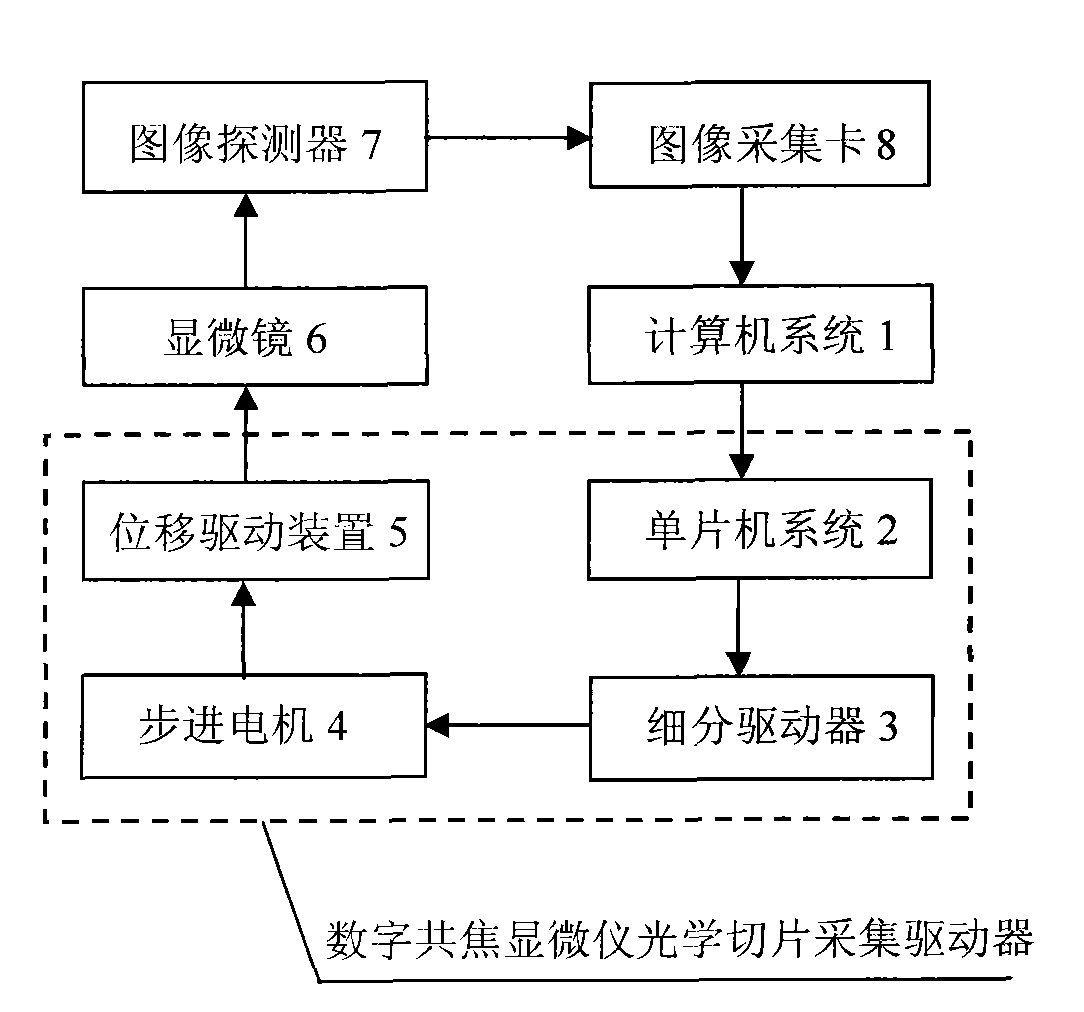
[0024] The optical slice acquisition driver of the present invention is composed of a single-chip microcomputer system 2, a subdivision driver 3, a stepping motor 4 and a displacement drive device 5, and is arranged on a microscope 6, controlled by the control software of the computer system 1, and adopts an image detector 7 Collect the serial optical section images of the biological cells imaged by the microscope 6 , convert them by the image acquisition card 8 and send them to the computer system 1 for display and storage. The computer system 1 has the functions of information sending, file operation, driver setting, image acquisition and image display according to the contents of the optical slice image acquisition method of the driver.

[0025] The working pr...


PUM


Abstract

The invention provides a driver of a digital confocal microscope optical section collecting device. The driver comprises a single-chip system, a sub-division driver, a stepping motor and a displacement driving device, and the driver arranged on the microscope and controlled by a computer drives a stage to acquire the sequenced optical section images of cells on the stage by an image detector. The method for the driver to acquire the optical section images is to carry out the submicron equidistant stepping control on the stage of the microscope by setting the section distance, collected section number and other parameters. By using the definition-based evaluation for the function value and the change rate thereof as the evaluation standards, the method of the invention is capable of automatically focusing on cells and locating the upper and lower edges, and acquiring the sequenced optical section images of intact cells.

Description

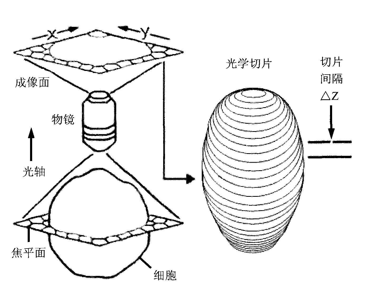
1. Technical field [0001] The invention relates to an optical slice collection driver for a digital confocal microscope. The driver is a component of a digital confocal microscope and belongs to the technical field of computer image acquisition and control. The driver is controlled by a computer to drive the stepwise displacement of the microscope stage at equal intervals, and uses the optical section image acquisition method of the driver to realize the purpose of sequential optical section acquisition of biological cells in the microscope. Background technique [0002] Since its birth, the biological optical microscope has provided a powerful tool for human beings to explore the microscopic world, and it is still widely used in various fields such as biomedical research. However, due to the optical diffraction effect and the defocused virtual image of the three-dimensional biological sample, the resolution is not high, the definition is low, and the details are difficult ...

Claims


Application Information

Patent Timeline
IPC IPC(8): G02B21/32G02B21/36
Inventor 陈华聂雄李江贺斌
Owner GUANGXI UNIV
Learn More