Medical digital subtraction image fusion method based on ridgelet transformation

A technology of ridgelet transform and digital subtraction, which is applied in the field of image fusion in medical image processing technology to achieve the effect of reducing the root mean square error rate, improving the efficiency of the algorithm, and being easy to implement.

Inactive Publication Date: 2010-08-11
SUZHOU UNIV
View PDF2 Cites 3 Cited by
  • Summary
  • Abstract
  • Description
  • Claims
  • Application Information

AI Technical Summary

Problems solved by technology

[0011] The purpose of the present invention is to provide a DSA image adaptive fusion method based on Ridgelet transform, in order to solve the shortcoming that the existing DSA image cannot realize adaptive fusion under the premise of keeping the details of blood vessel texture

Method used


Image

  • Medical digital subtraction image fusion method based on ridgelet transformation
  • Medical digital subtraction image fusion method based on ridgelet transformation
  • Medical digital subtraction image fusion method based on ridgelet transformation

Examples


Embodiment 1

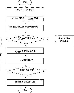
[0036] Embodiment one: figure 1 It is an overall flowchart of the DSA image adaptive fusion method based on Ridgelet transformation, and the data file (picture file) is a brain DSA blood vessel picture conforming to the DICOM standard.

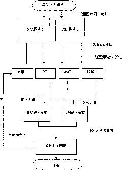
[0037] Step 1: Read in the DSA sequence picture and perform Ridgelet transformation on it. The specific steps are as follows (see figure 2 ):

[0038] Step 1: Read in two pictures in the DSA sequence picture, the picture size is uniformly set to 1024 pixels (length) × 1024 pixels (width) by the system, marked as picture 1 and picture 2. In this way, the problem of image distribution caused by too large images can be avoided.

[0039] Step 2: Carry out digital Ridgelet transformation on picture 1 and picture 2 respectively.

[0040] That is, through Radon transformation, the pixels of a 1024×1024 image become a 1024×2048 array, and then one-dimensional wavelet transformation is performed on the 1024×2048 array to obtain the Ridgelet transfor...


PUM


Abstract

The invention discloses a medical digital subtraction image fusion method based on ridgelet transformation, comprising the following steps of: (1) respectively carrying out the ridgelet transformation on two images to obtain ridgelet transformation matrixes; (2) fusing; (3) carrying out ridgelet inverse transformation on the fused ridgelet transformation matrixes to obtain a reconstructed image, i.e. a fused image. The invention is characterized in that in step (1), setting an initial judging threshold value and a step length for carrying out the ridgelet transformation, reconstructing the fused image by utilizing inverse transformation, calculating the information entropies of the fused image, changing the judging threshold value by using a dynamic fuzzy method according to the step length, repeating the operation, and then obtaining the ridgelet transformation matrixes in step (1) by using the judging threshold value corresponding to the maximum information entropy as the final judging threshold value. The invention can effectively improve the information entropies of the fused image, lower the error rates of mean square roots and has the performance better than that of other traditional fusion method. The algorithmic time has low complexity, the obtained result is more accurate, and therefore, the invention greatly enriches the grain details of medical images.

Description

technical field [0001] The invention belongs to the field of image fusion in medical image processing technology, and relates to an adaptive fusion method of medical digital subtraction (DSA) images, in particular to dynamic fuzzy technology and Ridgelet transformation theory. Background technique [0002] In today's world, cardiovascular and cerebrovascular diseases are seriously threatening human health and life safety. However, the diagnosis of vascular soft tissue is quite complicated, and relies heavily on high-tech methods such as computer and X-ray angiography. [0003] Digital Subtraction Angiography (DSA) is a new technology of medical imaging emerging in the 1980s. It is a new examination method combining computer and conventional X-ray angiography. It integrates digital electronics, computer technology and Angiography is a new technology integrating angiography. It has the characteristics of real-time imaging, high resolution, subtraction effect, and drawing of v...

Claims


Application Information

Patent Timeline
IPC IPC(8): G06T5/50
Inventor 崔志明张广铭吴健
Owner SUZHOU UNIV
Why Eureka
Try Eureka