Dynamic three-dimensional reconstruction method of single-arm X-ray angiogram maps

A technology for three-dimensional reconstruction and angiography, which is applied in image analysis, image data processing, and instruments used for radiological diagnosis, etc., can solve the problem of not taking into account the specificity of cardiac motion, and achieve the effect of improving reconstruction accuracy and overcoming limitations.

Active Publication Date: 2010-08-11
HUAZHONG UNIV OF SCI & TECH
View PDF1 Cites 16 Cited by
  • Summary
  • Abstract
  • Description
  • Claims
  • Application Information

AI Technical Summary

Problems solved by technology

The method is to directly use the motion vector of the blood vessel point in the model projection map as the compensation vector o

Method used


Image

  • Dynamic three-dimensional reconstruction method of single-arm X-ray angiogram maps
  • Dynamic three-dimensional reconstruction method of single-arm X-ray angiogram maps
  • Dynamic three-dimensional reconstruction method of single-arm X-ray angiogram maps

Examples


Embodiment Construction

[0048] The present invention will be further described below in conjunction with the drawings and specific embodiments:

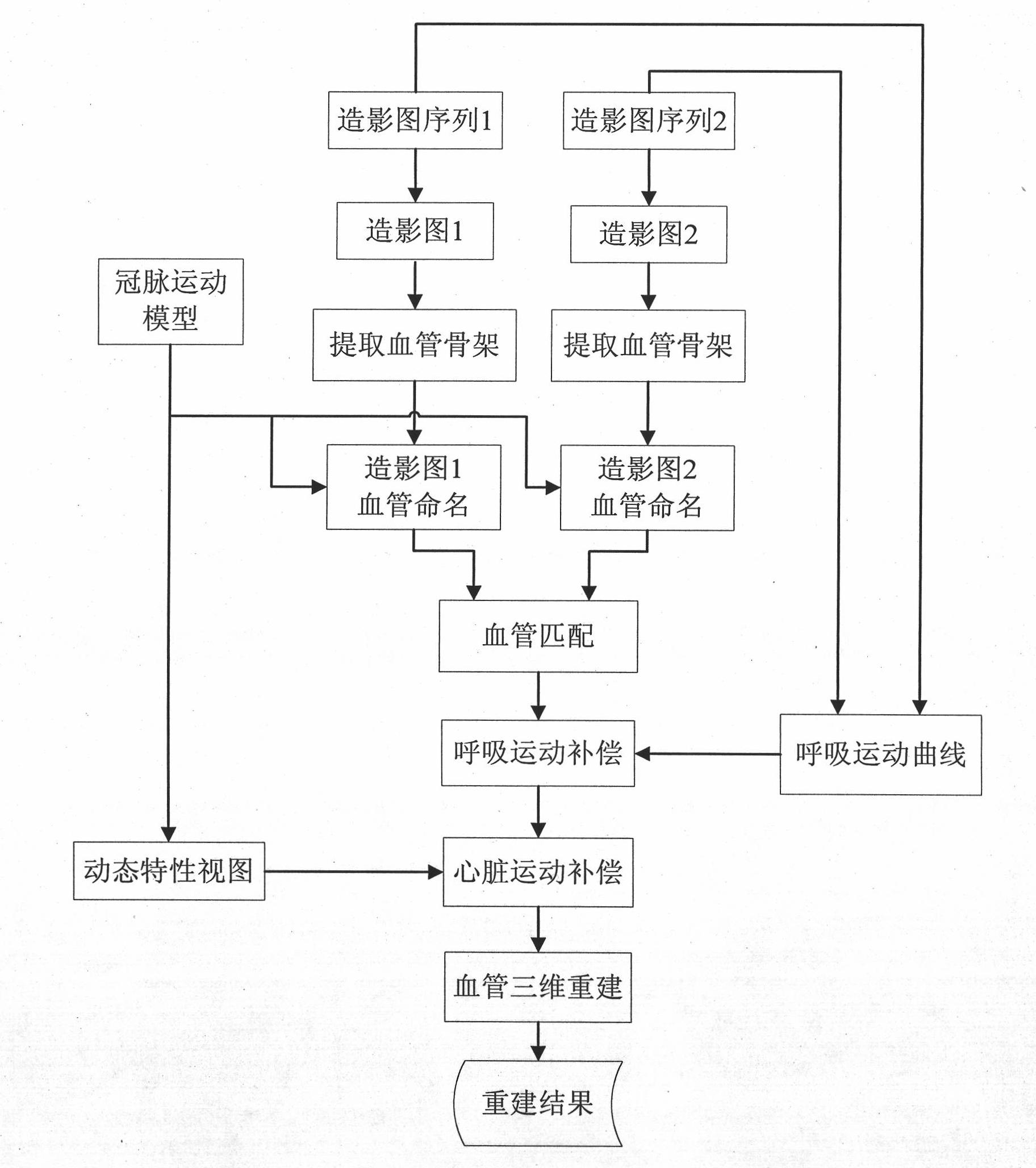
[0049] The invention uses the Fourier series expansion method to extract the respiratory motion curve to perform respiratory motion compensation on the two-dimensional coronary angiogram, and uses the motion characteristics constructed by the coronary motion model to try to guide the heart motion compensation, so that two single-arm X-ray imaging The images are in the same phase of the heart and breathing movement, so that three-dimensional reconstruction can be performed. figure 1 It is a flowchart of the method of the present invention, and the specific steps are as follows:

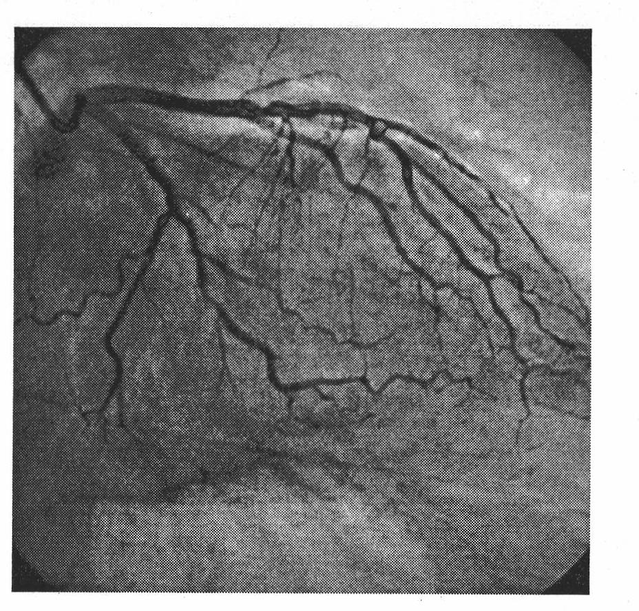
[0050] (1) Extraction of blood vessel topology from X-ray imaging

[0051] The X-ray image is segmented to obtain the skeleton of the coronary blood vessel, and the eight-connected chain code is used to track the cardiovascular skeleton, the radius of the blood vessel is extracted, and th...


PUM


Abstract

The invention relates to a dynamic three-dimensional reconstruction method of single-arm X-ray angiogram maps, which belongs to the field of the intersection of digital image processing and medical imaging and aims to meet the requirements of the auxiliary detection and surgical navigation of cardiovascular diseases in clinical medicine. The invention provides a concept of 'dynamic vessel three-dimensional reconstruction', so that the angiogram maps of double visual angles at different moments are compensated for respiratory movement and cardiac movement. The dynamic three-dimensional reconstruction method can obtain the high three-dimensional reconstruction accuracy of coronary artery vessels, solve the problem that the automatic and reliable three-dimensional reconstruction is performed by multi-visual angle angiography maps with different time phases, assist in the detection and surgical navigation of the cardiovascular diseases effectively and meet the clinical requirement.

Description

Technical field [0001] The invention belongs to the intersecting field of digital image processing and medical imaging, and specifically relates to a three-dimensional reconstruction method of a single-arm X-ray contrast image. Background technique [0002] The three-dimensional reconstruction of the vascular tree is a process of restoring the three-dimensional structure of the blood vessel through the corresponding image information in the X-ray two-dimensional projection images from different perspectives. It is very different from ordinary visible light image 3D reconstruction. X-ray imaging is an image formed on a fluorescent screen after the X-rays projected to the human body are attenuated by different tissues in the human body. The value of each pixel is the superposition of the attenuation of all tissues on the X-ray path. The background is complex and noise Very strong, it is very difficult to reconstruct the three-dimensional cardiovascular tree from the X-ray image. ...

Claims


Application Information

Patent Timeline
IPC IPC(8): G06T17/00G06T7/20A61B6/00G06T7/223
Inventor 张天序邓觐鹏孙祥平肖晶黎云曹治国桑农王国铸王芳
Owner HUAZHONG UNIV OF SCI & TECH
Why Eureka
Try Eureka