
Method and system for imaging vessels

A technology of vascular imaging and blood vessels, which is applied in the field of vascular imaging, can solve problems such as muscle and bone injuries, hands and arms of the sonographer, hindering the use of transcranial Doppler examination, etc., to achieve the effect of eliminating pressure and injury

Inactive Publication Date: 2010-11-17
KONINK PHILIPS ELECTRONICS NV
View PDF1 Cites 17 Cited by
  • Summary
  • Abstract
  • Description
  • Claims
  • Application Information

AI Technical Summary

Problems solved by technology

Training and practice to master these techniques may preclude use of transcranial Doppler
Additionally, the uncomfortable and strenuous positioning of the sonographer's hand and arm during the examination can lead to musculoskeletal injury

Method used


Image

  • Method and system for imaging vessels
  • Method and system for imaging vessels
  • Method and system for imaging vessels

Examples


Embodiment Construction

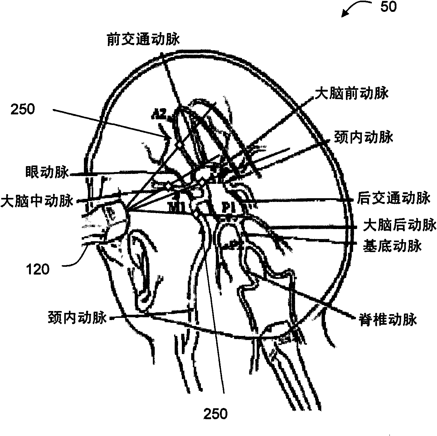
[0016] Exemplary embodiments of the present invention are described with respect to data capture, imaging of blood vessels and mapping of blood flow for transcranial Doppler examination of the circle of Willis in humans. Those of ordinary skill in the art will appreciate that the exemplary embodiments of the present invention may be applied to blood vessels in other parts of the body, whether human or animal. Using the methods and systems of exemplary embodiments of the present invention, the methods and systems of the exemplary embodiments of the present invention can Suitable for application to vessels other than the Circle of Willis.

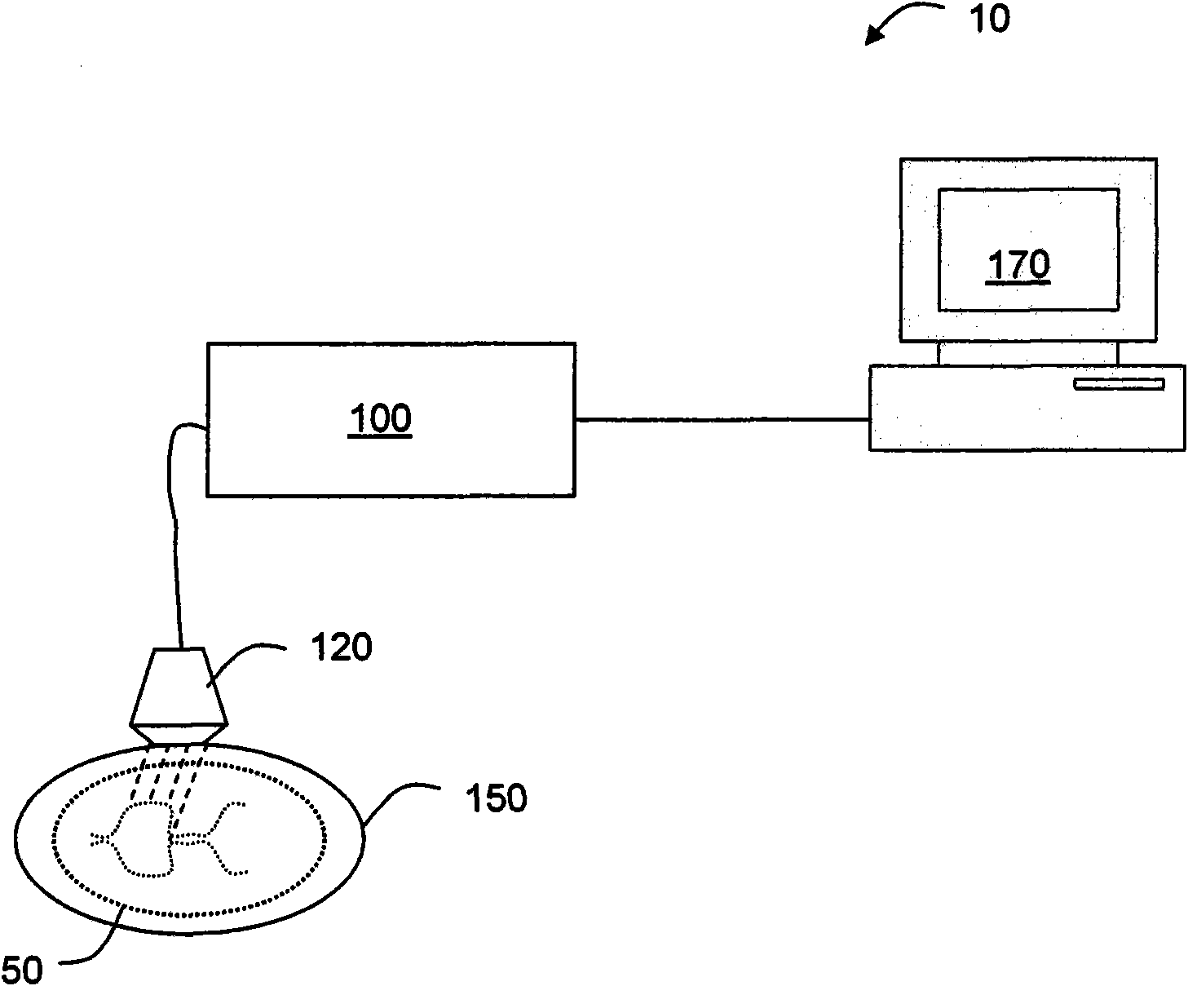
[0017] With reference to the drawings and in particular to figure 1 , which shows an ultrasound imaging system according to an exemplary embodiment of the present invention, and which is generally indicated by reference numeral 10 . System 10 may perform ultrasound imaging on a patient's head 50 and may include a processor or other control ...


PUM


Abstract

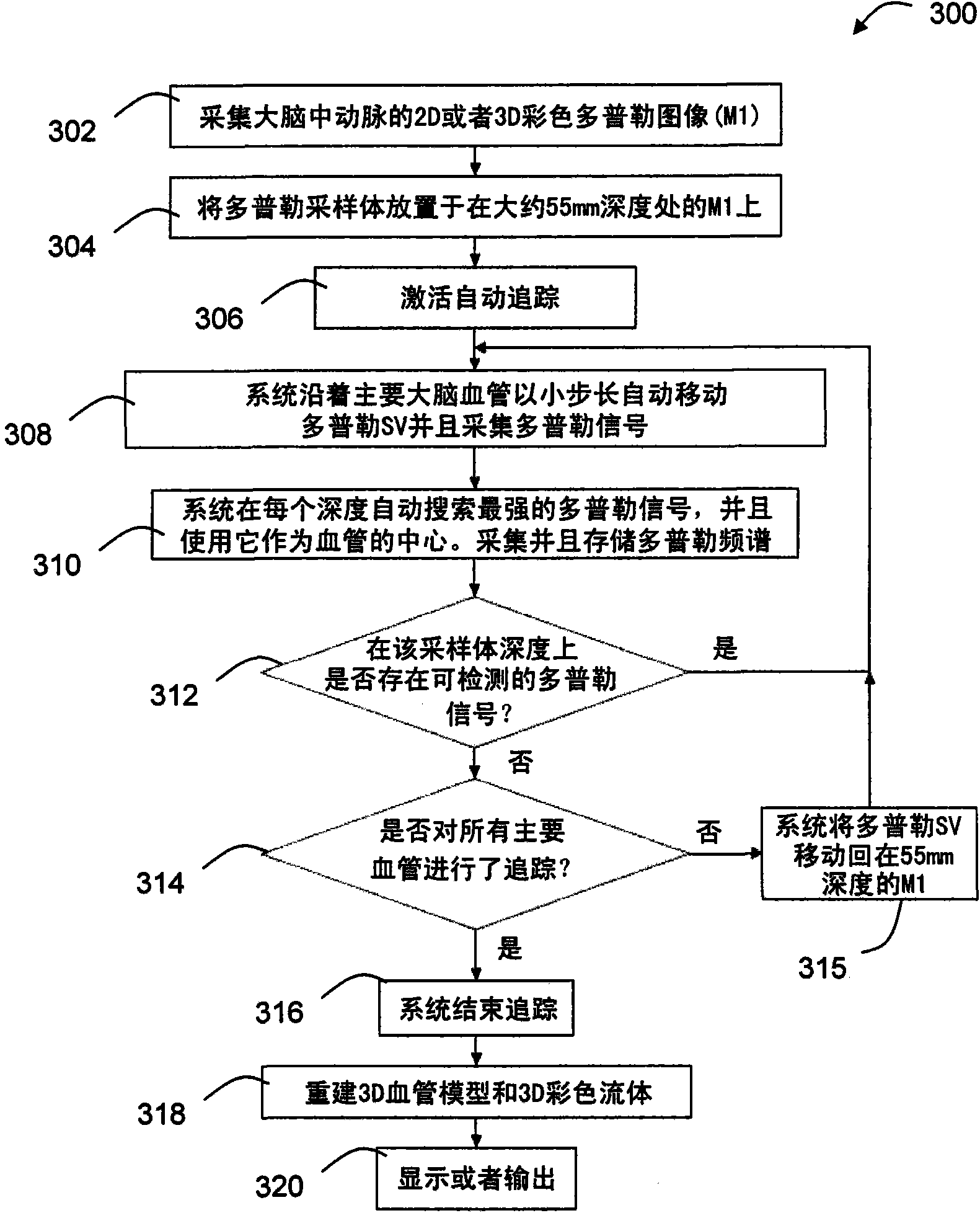
A method and system of imaging of a vessel is provided. The system can include a matrix transducer array (120) for transmitting ultrasonic waves into a region of a body having a vessel and receiving echoes in response, where the echoes are associated with blood flow through the vessel; and a processor (100) operably coupled to the matrix transducer array. The processor can adjust positions of sample volumes (250) associated with the echoes. The processor can electronically steers the ultrasonic waves at one or more of the positions of the sample volumes. The processor can determine a wall of the vessel at each of the positions of the sample volumes based on a Doppler spectrum captured at each of the positions of the sample volumes. Other embodiments are disclosed.

Description

technical field [0001] The present invention relates generally to imaging systems, and more particularly to methods and systems for imaging blood vessels. Background technique [0002] Imaging blood vessels may be based on detecting blood flow through the blood vessels. For example, transcranial Doppler is a test that measures the velocity of blood flow through blood vessels in the brain to diagnose subarachnoid hemorrhage, among other problems, such as thrombosis, stenosis, vasospasm. [0003] Transcranial Doppler can be performed with "B-mode" imaging, which displays a two-dimensional image of what the ultrasound probe sees. Once the operator is able to locate the desired vessel, blood flow velocity can be measured using a pulsed Doppler probe, which plots the velocity versus time. Together these form a bidirectional test. The second recording method may use only the second probe function, and may also rely on the operator's training and experience to find the correct v...

Claims


Application Information

Patent Timeline
Patent Type & Authority Applications(China)
IPC IPC(8): G01S15/89G01S7/52A61B8/06G10K11/34
CPCA61B8/4483A61B8/42A61B8/06G01S15/8993A61B8/483G01S7/52073G01S15/8979A61B8/0816
Inventor X·史
Owner KONINK PHILIPS ELECTRONICS NV
Learn More