Method for retrospectively classifying chest or abdomen computed tomography (CT) images based on respiratory phase

A CT image and respiratory phase technology, applied in the field of medical CT image processing, can solve the problems of inaccuracy, affecting the effect of radiotherapy, unable to reconstruct 4D-CT normally, and achieve the effect of improving accuracy

Inactive Publication Date: 2011-05-25
SOUTHERN MEDICAL UNIVERSITY
View PDF3 Cites 21 Cited by
  • Summary
  • Abstract
  • Description
  • Claims
  • Application Information

AI Technical Summary

Problems solved by technology

Since the computerized tomography imaging equipment with the PRM system has a set of optical system (PRM system) to obtain two fluorescent marker points, we don't talk about its complexity and high equipment cost. It is said that there are also following defects: 1, because the CT scan and the execution of the plan of tumor radiotherapy are often not carried out on the same bed, so there is inevitably an error of changing the bed; 2, even if it is carried out on the same bed, due to the The plastic box with fluorescent markers in the optical syst

Method used


Image

  • Method for retrospectively classifying chest or abdomen computed tomography (CT) images based on respiratory phase
  • Method for retrospectively classifying chest or abdomen computed tomography (CT) images based on respiratory phase
  • Method for retrospectively classifying chest or abdomen computed tomography (CT) images based on respiratory phase

Examples


Embodiment Construction

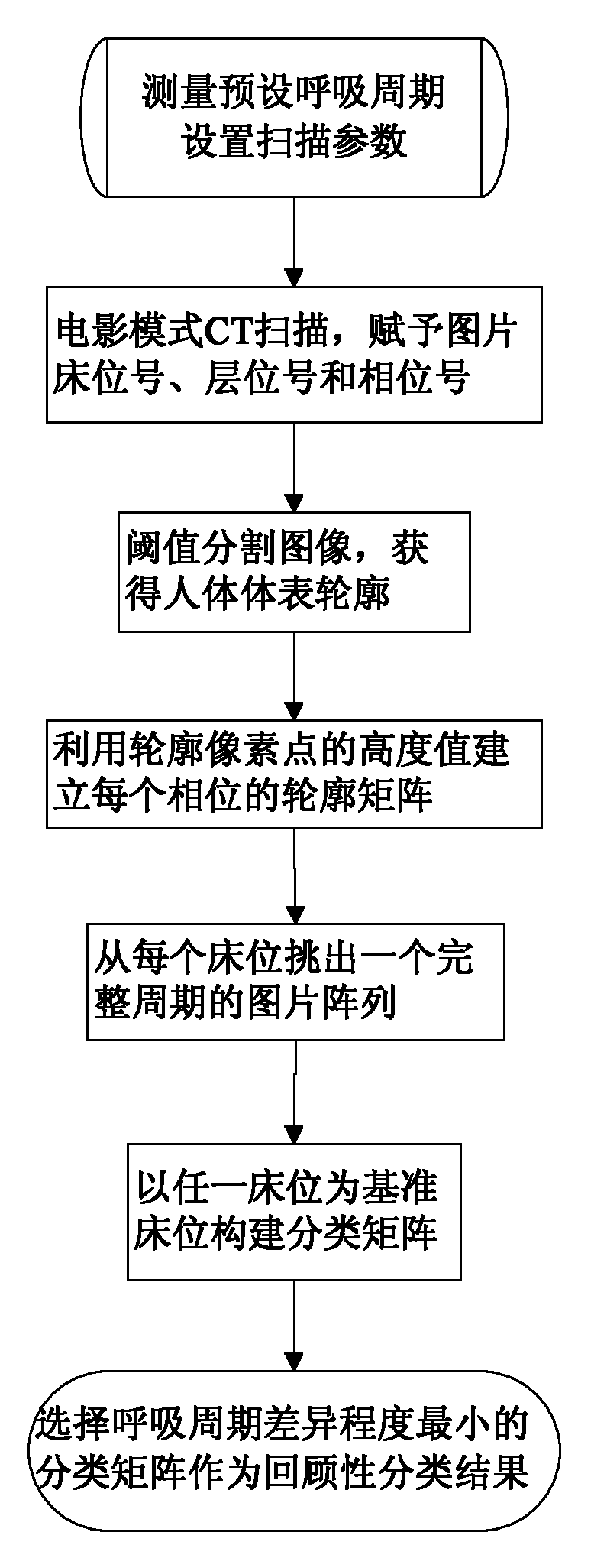
[0025] The implementation object of this example is a patient suffering from lung cancer. The CT scanner used is GE lightspeed 16-slice CT. The duration of each revolution of the CT machine is 0.5 seconds, and the size of each picture is 512×512. Such as figure 1 As shown, the retrospective classification process of the CT images of the patient suffering from lung cancer is as follows:

[0026] (1) Let the patient lie on the CT bed first, use the ventilator to measure the average breathing cycle of 5 seconds, and use this as the preset breathing cycle, and then draw up the scanning plan as follows: scan 3 beds, each bed has 8 layers , the thickness of each layer is 1.5mm, the CT scanner rotates 10 times for each preset breathing cycle, and the scanner rotates 16 times for each bed, and scans in cine mode.

[0027] (2) Start the CT scanner to perform cine-mode CT scanning on the chest of the human body, and assign bed numbers and layer numbers to each of the obtained CT pictur...


PUM


Abstract

The invention relates to a method for retrospectively classifying chest or abdomen computed tomography (CT) images based on a respiratory phase. The method comprises the following steps of: measuring a preset respiratory cycle and setting parameters of CT scanning; performing movie-mode CT scanning so as to obtain images and endowing each image with a bed number, a layer number and a phase number; performing threshold value segmentation on each image so as to obtain the surface profile of a human body; establishing a profile matrix of each phase by using the height values of pixel points of the surface profile of the human body; accumulating the height values of elements in the profile matrix of each phase and picking out a group of images which correspond to a maximum accumulated sum value from each bed, wherein the group of images serve as an image array of a complete cycle; performing cubic spline smooth fitting on each row of vectors of a profile matrix between two phases by taking any bed as a reference bed, calculating the distance sum of profile height difference between adjacent layers of two phases and picking out image sequence numbers with the minimum distance sum so as to establish a classification matrix; and selecting a classification matrix with the minimum retrospective cycle difference degree, wherein the classification matrix is taken as a retrospective classification result.

Description

technical field [0001] The invention relates to a method for processing medical CT images, in particular to a method for classifying human CT images according to respiratory phases, and the obtained CT images can be used to formulate tumor radiotherapy plans. Background technique [0002] Computed tomography, or CT (Computed Tomography), is an essential tool and technology for tumor radiation therapy. 4D-CT is a new technology to evaluate the movement of tumors and organs. During the CT scanning process, the time information representing the patient's respiratory movement is recorded synchronously. Therefore, in addition to the three-dimensional spatial information, the scanning results also include the patient's respiratory movement. time information. It can realize the dynamic observation of the shape of the moving tumor, and provides conditions for studying the movement of the tumor. Compared with 3D-CT, 4D-CT can not only reduce the motion artifacts of organs but also ...

Claims


Application Information

Patent Timeline
IPC IPC(8): A61B6/03
Inventor 周凌宏徐圆卢文婷刘迎军甄鑫
Owner SOUTHERN MEDICAL UNIVERSITY
Why Eureka
Try Eureka