Eureka AIR delivers breakthrough ideas for toughest innovation challenges, trusted by R&D personnel around the world.

Biological digital microscope with double ccd (charge coupled device) light sensitive elements and photographic image processing method thereof

A digital microscope and photosensitive element technology, applied in the field of digital microscopes, can solve the problems of tired efficiency, lack of objectivity and standardization, lack of social medical resources, pathologist resources, etc., and achieve the effect of improving accuracy

Active Publication Date: 2011-08-10
南京福怡科技发展股份有限公司
View PDF4 Cites 13 Cited by
  • Summary
  • Abstract
  • Description
  • Claims
  • Application Information

AI Technical Summary

Problems solved by technology

At the same time, since pathological diagnosis is based on the experience of doctors, the so-called gold standard of pathological diagnosis is also based on the premise of individual experience, which is not objective and standardized.
Also working under the microscope for long hours is tiring and inefficient
In addition, the lack of a large number of pathologists in medical institutions has also resulted in an extreme shortage of social medical resources, especially pathologist resources.

Method used


Image

  • Biological digital microscope with double ccd (charge coupled device) light sensitive elements and photographic image processing method thereof
  • Biological digital microscope with double ccd (charge coupled device) light sensitive elements and photographic image processing method thereof
  • Biological digital microscope with double ccd (charge coupled device) light sensitive elements and photographic image processing method thereof

Examples


Embodiment Construction

[0015] In order to enable the public to fully understand the technical essence and beneficial effects of the present invention, the specific embodiments of the present invention will be described in detail below in conjunction with the accompanying drawings, but the applicant's description of the embodiments is not a limitation to the technical solution. Changes in form rather than in substance should be regarded as the protection scope of the present invention.

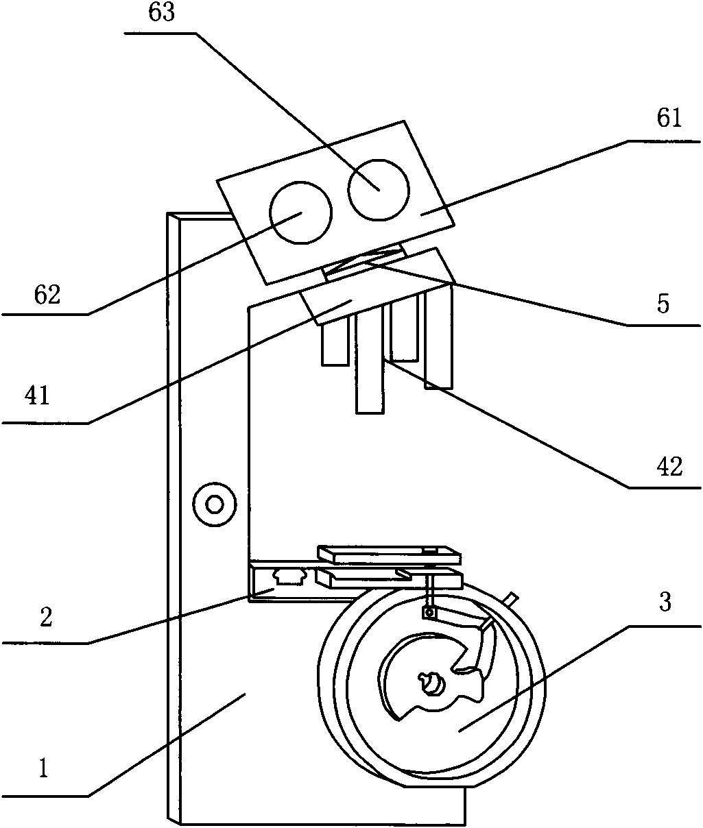
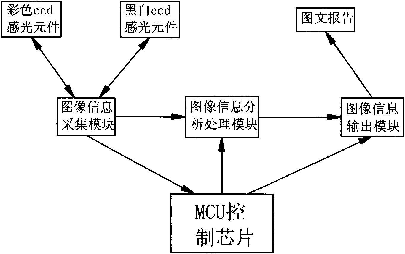
[0016] Such as figure 1 with figure 2As shown, the biological digital microscope with dual ccd photosensitive elements of the present invention includes a frame 1 and a microscopic platform 2 respectively installed on the frame 1, an MCU control chip, a lens barrel 61, an objective lens conversion seat 41, and is used for scanning A barcode scanning device for encoding information on the slide glass and a specimen image information acquisition device for collecting image information of the slide glass, the specimen...


PUM


Abstract

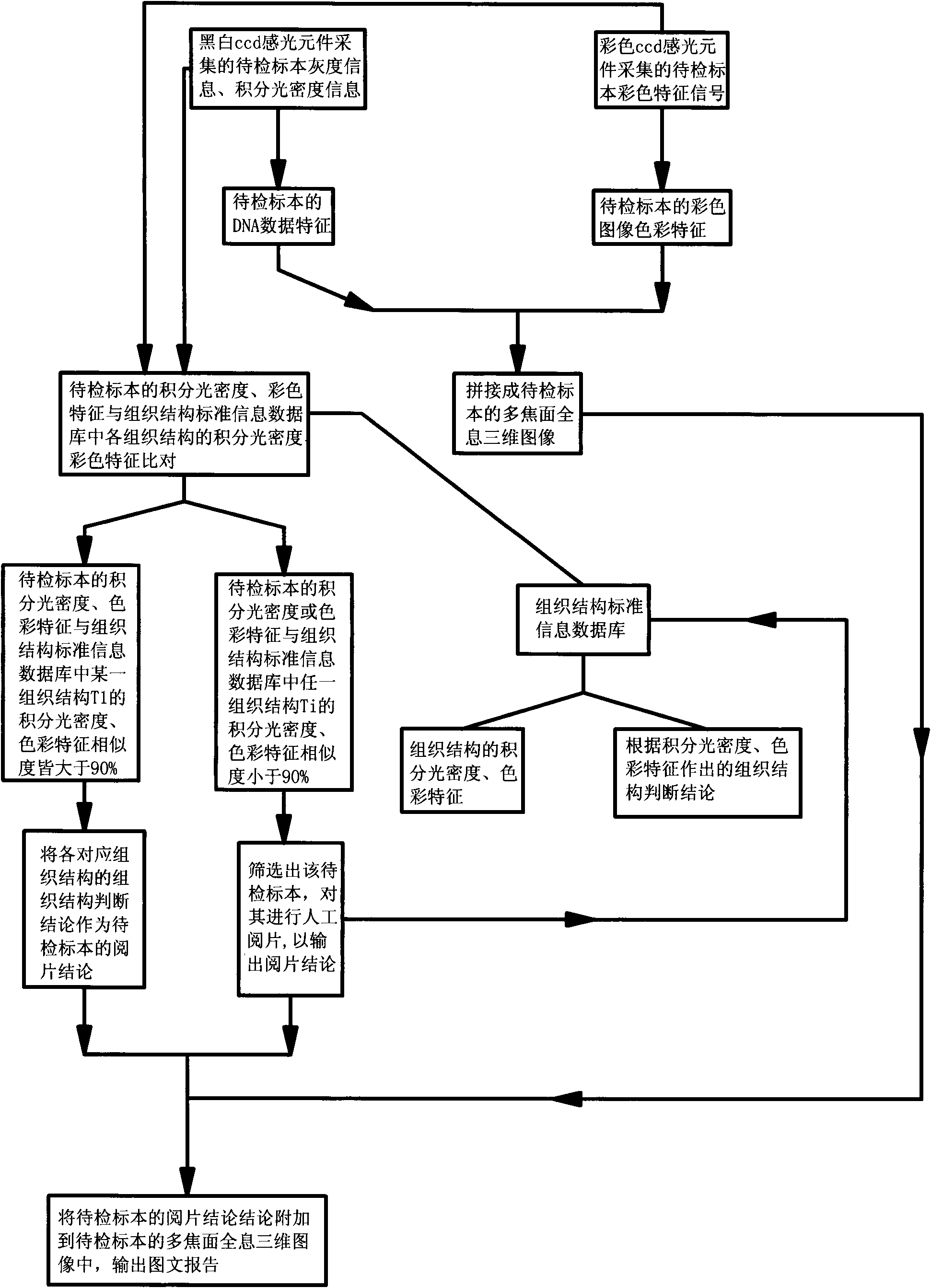
The invention discloses a biological digital microscope with double ccd (charge coupled device) light sensitive elements and a photographic image processing method thereof, wherein a colour ccd light sensitive element and a black and white ccd light sensitive element are used for collecting image information; the black and white ccd light sensitive element collects a gray scale and integrating optical density information of a specimen cell nucleus; the colour ccd light sensitive element collects a colour characteristic signal of a specimen image; the three-dimensional split joint of a specimen picture is performed by the gray scale of a specimen cell nucleus, the integrating optical density information and colour characteristic signal of a specimen image, so as to form a multi-focal plane holographic three-dimensional image; simultaneously, a film reading conclusion is output according to the integrating optical density information of a cell nucleus; finally a graphics context report of the specimen to be examined is output; therefore the biological digital microscope disclosed by the invention can perform conventional morphological observation for the specimen, as well as can perform digital photographing for the dyed DNA (deoxyribonucleic acid) for forming a multi-focal plane holographic three-dimensional image which is convenient for observing all the information of the cell in a multilayer and multi-focal plane mode; and the accuracy of early diagnosis is greatly improved.

Description

technical field [0001] The invention relates to a digital microscope, in particular to a biological digital microscope with double ccd photosensitive elements and a photographic image processing method thereof, which are mainly used in microscopic examination of tissue sections stained by HE-Feulgen. Background technique [0002] The development of medicine, especially the development of pathology, mainly relies on the magnification of optical microscopes to provide a hardware basis for doctors and researchers to observe tissue samples in detail. In the field of pathology research as well as in everyday medical practice, there is a growing need for the processing of complex data, the application of record-keeping or archiving systems, and the possibility of interactive consultation of examination results. [0003] At present, the most advanced microscope simply adds a camera interface outside the eyepiece, which plays the role of collecting images and putting them on the dis...

Claims


Application Information

Patent Timeline
Patent Type & Authority Applications(China)
IPC IPC(8): G02B21/36G01N21/17G06T17/00G06K9/62G06F17/30
Inventor 姚斌
Owner 南京福怡科技发展股份有限公司
Why Eureka
Eureka Blog
Learn More