
Biological information imaging apparatus

A technology for biological information and imaging devices, which can be used in measurement devices, ultrasonic/sonic/infrasound image/data processing, and diagnosis using light, and can solve problems such as individual differences

Inactive Publication Date: 2011-08-10
CANON KK
View PDF2 Cites 12 Cited by
  • Summary
  • Abstract
  • Description
  • Claims
  • Application Information

AI Technical Summary

Problems solved by technology

Although the average effective attenuation coefficient μ of the living body eff It is already known about some parts, but the effective attenuation coefficient μ eff There are individual differences

Method used


Image

  • Biological information imaging apparatus
  • Biological information imaging apparatus
  • Biological information imaging apparatus

Examples


no. 1 example

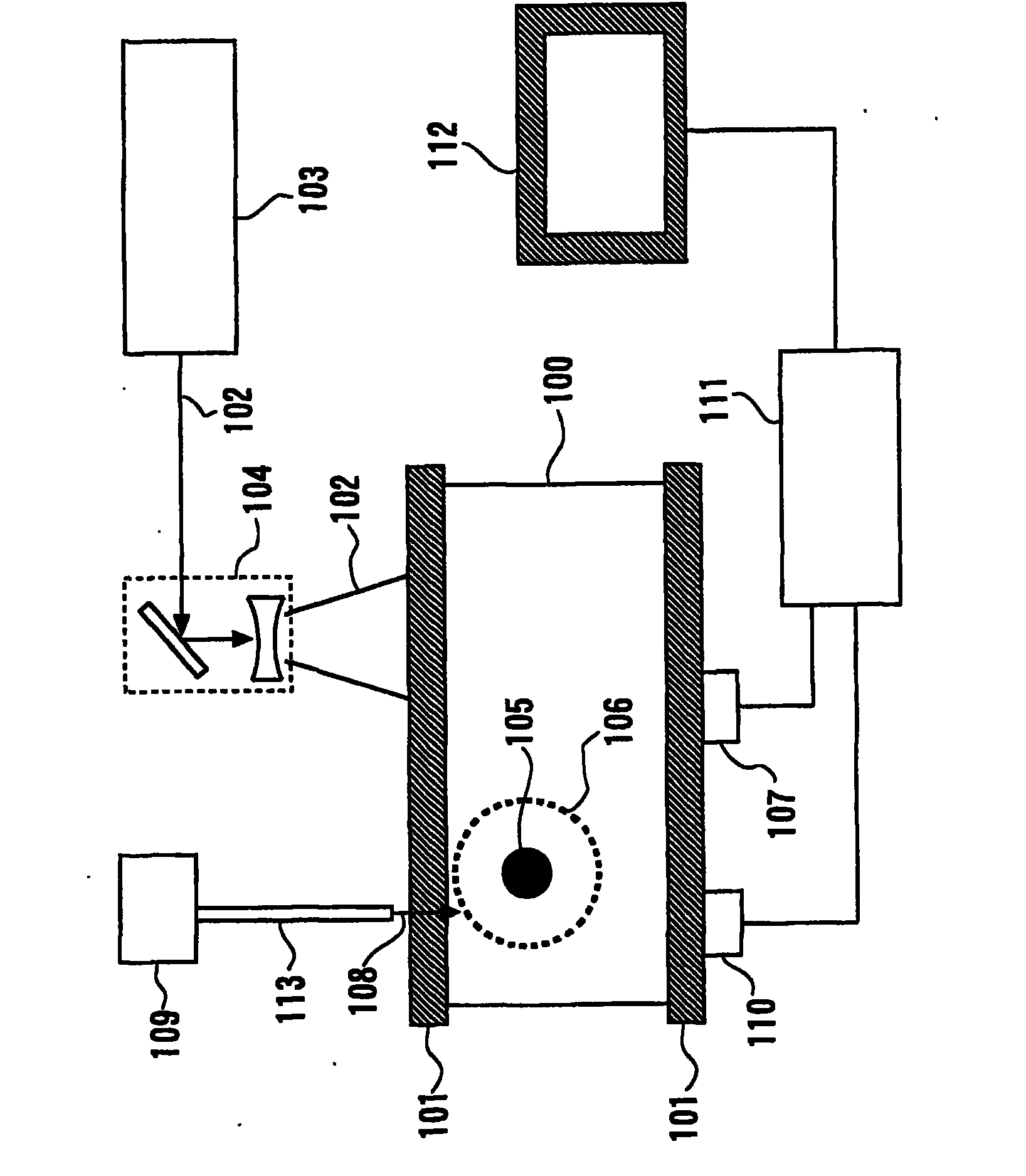
[0040] figure 1 A biological information imaging device according to a first embodiment of the present invention is shown. The biological information imaging apparatus described in this embodiment is capable of displaying the distribution of optical properties of a living body and the concentration distribution of substances constituting biological tissues obtained from the information as images in order to diagnose malignant tumors or diseases in blood vessels or to observe the progress of chemotherapy device.

[0041] In the biological information imaging apparatus according to the present embodiment, the subject 100 as a living body is sandwiched and fixed between two fixing members 101 . In addition, first light 102 irradiated from a first light source 103 is guided into the subject 100 via an optical unit 104 constructed with a lens or the like to irradiate the subject 100 . At this time, the energy of the first light 102 is absorbed by a light absorber 105 such as a bl...

no. 2 example

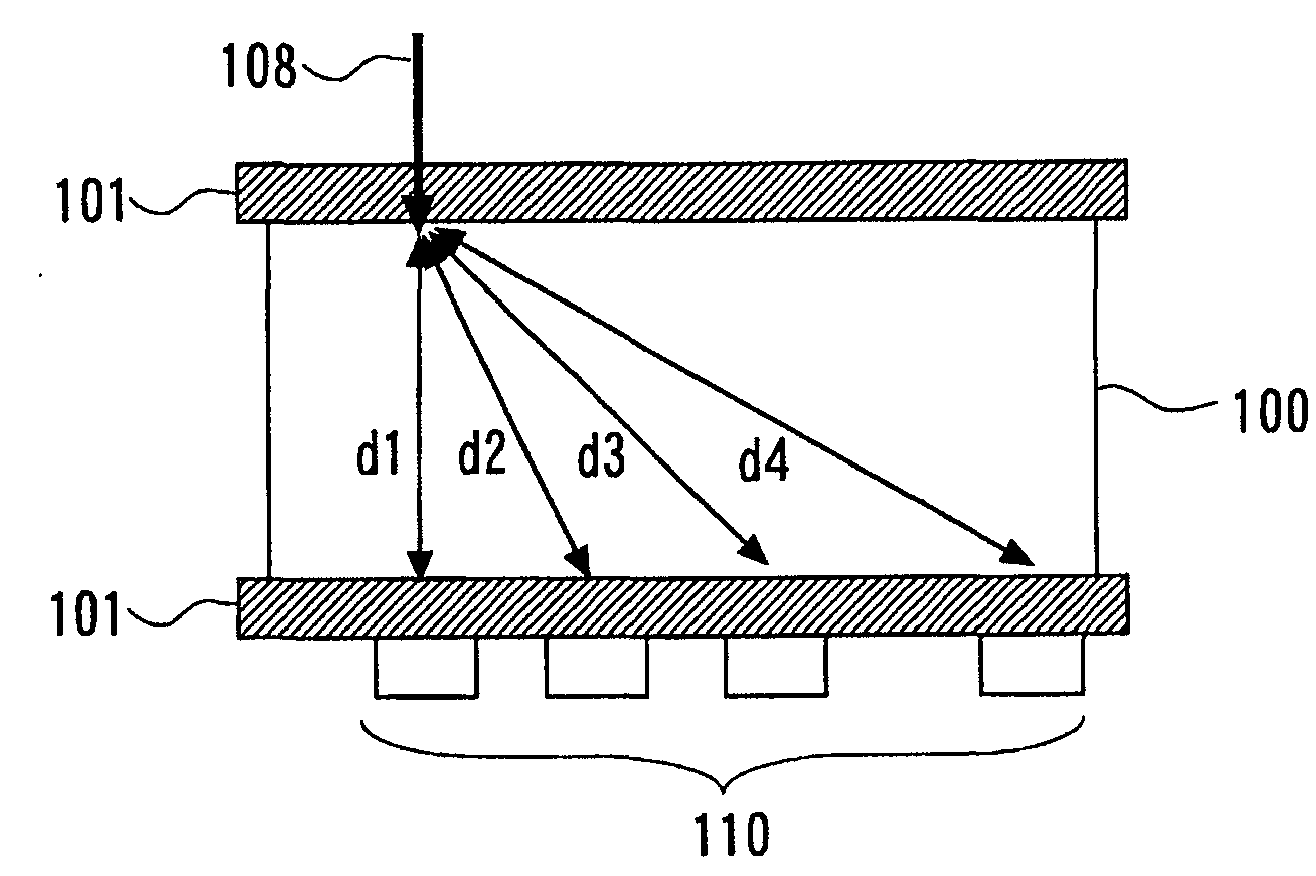
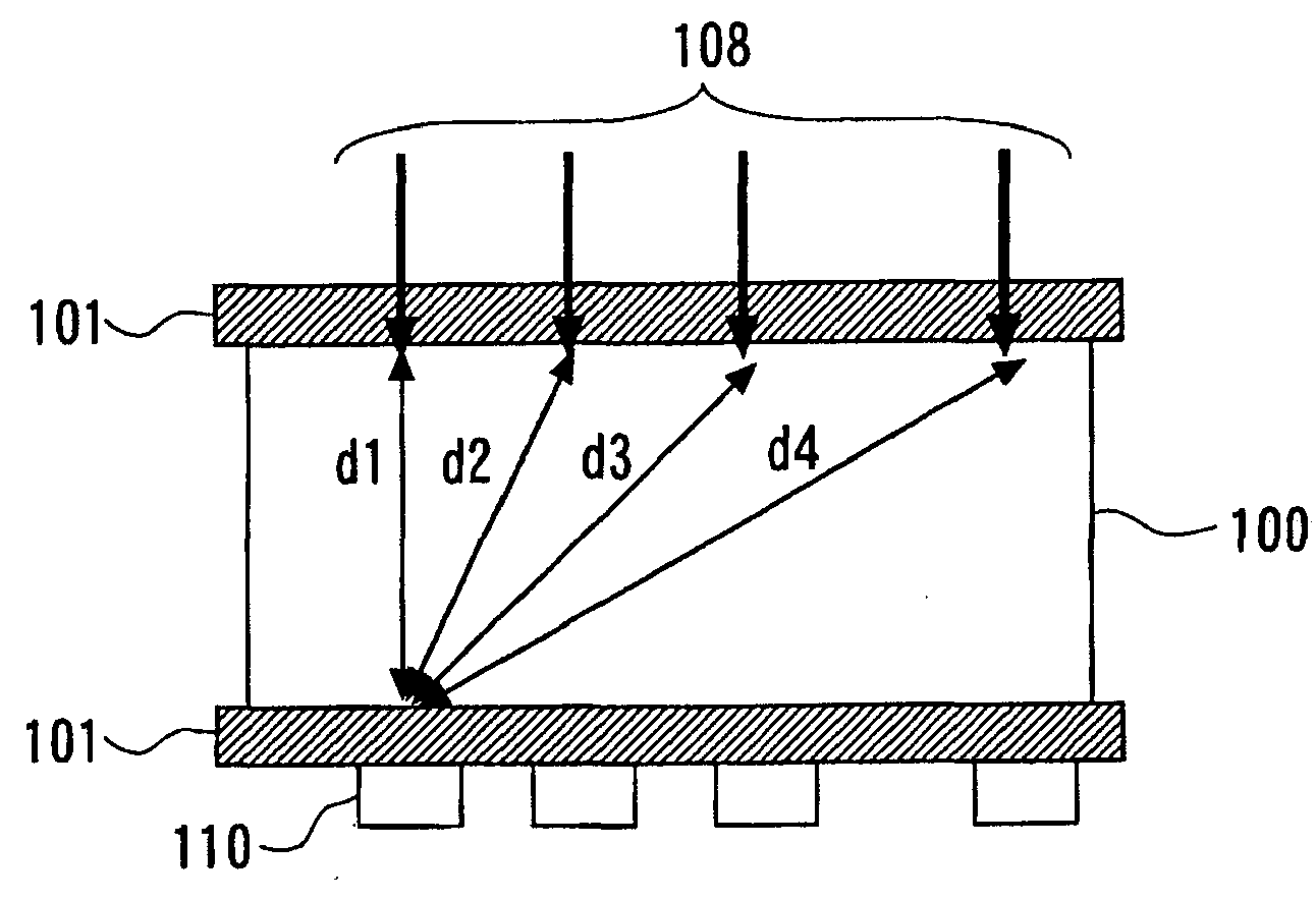
[0080] A second embodiment of the present invention will now be described with reference to the accompanying drawings. In this embodiment, the second electrical signal is also detected by the first light 102 to obtain the effective attenuation coefficient μ eff example of. Figure 7 The biological information imaging device according to the present embodiment is shown. The biological information imaging device according to the present embodiment differs from that of the first embodiment in that the second light source 109 and the optical waveguide 113 are not included, and the second light 108 is not used. In the following, the same components as those of the first embodiment are denoted by the same reference numerals, and descriptions thereof will not be repeated. Only features different from those of the first embodiment are described.

[0081] In this embodiment, the second electric signal obtained by detecting the first light 102 irradiated from the first light source 1...


PUM


Abstract

The biological information imaging apparatus includes an acoustic wave detector 107 that detects an acoustic wave that is generated from a light absorber 105 and converts it to a first electrical signal; a photo-detector 110 that detects intensities of the light corresponding to a plurality of propagation distances of the light which propagates through the specimen 110 and converts it to a second electrical signal; a signal processing apparatus 111 that derives an average effective attenuation coefficient [mu]eff of the specimen 110 based on the second electrical signal and derives an optical absorption coefficient [mu]a of the specimen 110 based on the first electrical signal and the average effective attenuation coefficient [mu]eff; and an image constructing apparatus 111 that constructs an image of the distribution of the optical absorption coefficient [mu]a based on the distribution of the optical absorption coefficient [mu]a.

Description

technical field [0001] The invention relates to a biological information imaging device. Background technique [0002] In recent years, biological information imaging devices for acquiring images from information of a living body by using techniques such as X-rays, ultrasonic waves, and magnetic resonance imaging (MRI) techniques have been widely used in the medical field. In addition, an optical imaging device in which light irradiated from a light source such as a laser is allowed to propagate in a living body or the like and the propagated light or the like is detected has been actively studied in the medical field. in order to obtain information about living organisms. As one of optical imaging techniques, photoacoustic tomography (PAT) has been proposed (for example, refer to Non-Patent Document 1). [0003] In photoacoustic tomography, pulsed light generated from a light source is irradiated to a living body as a subject, and acoustic waves generated from biological ...

Claims


Application Information

Patent Timeline
Patent Type & Authority Applications(China)
IPC IPC(8): A61B5/00A61B8/00A61B8/08A61B8/13G01N21/17
CPCA61B5/0059G01N2201/0662A61B5/0095A61B5/02007G01N21/1702
Inventor 中嶌隆夫福谷和彦
Owner CANON KK
Learn More