X-ray computerized tomography system and method

A technology of tomography and computer, which is applied in the field of medical imaging, can solve the problems of high cost and difficult implementation, and achieve the effects of easy implementation, reduction of X-ray dose, and cost reduction

Active Publication Date: 2012-04-04
SIEMENS SHANGHAI MEDICAL EQUIP LTD
View PDF5 Cites 16 Cited by
  • Summary
  • Abstract
  • Description
  • Claims
  • Application Information

AI Technical Summary

Problems solved by technology

The former usually needs to greatly change the existing algorithm of the CT system, while the lat...

Method used


Image

  • X-ray computerized tomography system and method
  • X-ray computerized tomography system and method
  • X-ray computerized tomography system and method

Examples


Embodiment Construction

[0030] In order to make the purpose, technical solution and advantages of the present invention clearer, the following examples are given to further describe the present invention in detail.

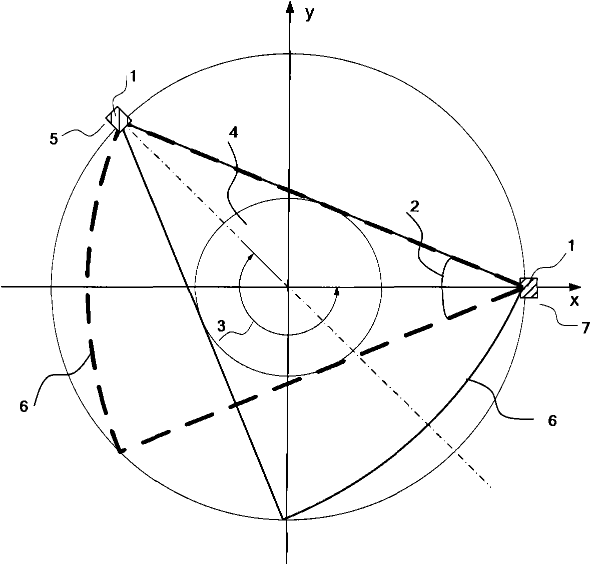
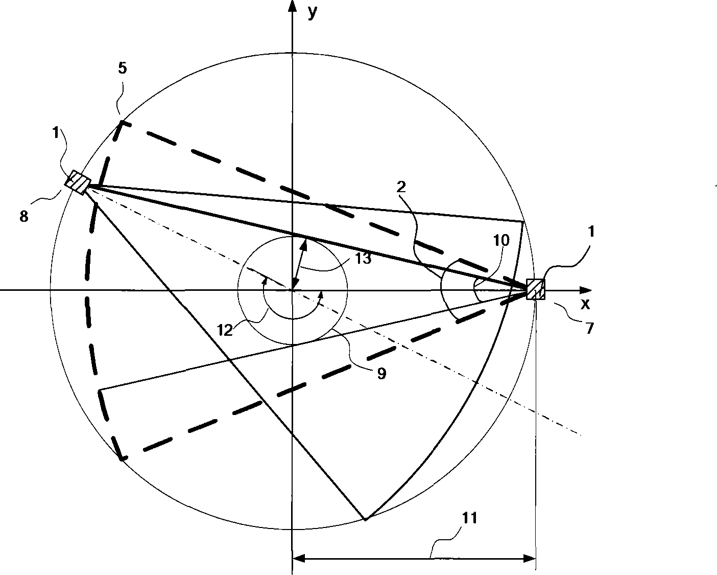
[0031] In the present invention, the object to be inspected may be a certain area of ​​the human body, or a certain organ or tissue of the human body. The scanning direction is the direction in which the examination bed enters and exits the rack in the CT system, usually the z direction.

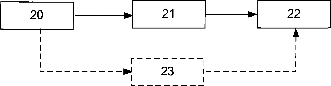
[0032] The CT scanning system of the present invention is used for partially scanning the object to be checked, such as image 3 Shown is a schematic diagram of the components of the CT scanning system of the present invention, including: an intermediate angle calculation component 20, a reconstruction angle calculation component 21 and a scanner 22, wherein the intermediate angle calculation component 20 is used to calculate , FOV) cross-section size and center coordinates to calculate the intermedia...


PUM


Abstract

The invention discloses an X-ray computerized tomography system, which is used for partially scanning an object to be detected. The system comprises a middle angle computing module, a rebuilding angle computing module and a scanner, wherein the middle angle computing module is used for computing a middle angle according to the size of the vision field cross section and the central coordinate of the object to be detected, and transmitting the middle angle to the rebuilding angle computing module; the rebuilding angle computing module is used for adding the middle angle to 180 degrees to obtain a rebuilding angle, and transmitting the rebuilding angle to the scanner; and the scanner is used for scanning a part of the object to be detected according to the rebuilding angle. The invention further discloses an X-ray computerized tomography method. Due to the adoption of the system and the method, the scanning time can be shortened, the time resolution can be increased, and the radiation dosage received by a patient can be reduced.

Description

technical field [0001] The invention relates to the field of medical imaging, in particular to an X-ray computed tomography system and method. Background technique [0002] In X-ray computed tomography (Computed Tomography, CT), in order to obtain high temporal resolution, dual-source CT and partial scanning are often used. Partial scans are between half scans and full scans, and are advantageous when scanning organs or tissues in motion (such as the heart), because the shorter the scan time, the less likely the object to be examined is to move during the imaging process, thereby reducing The impact of motion artifacts on image quality was investigated. [0003] Part of the scan has a rotational scan angle of less than 360°. At present, in partial scans of CT systems, the scan time is usually determined according to a fixed reconstruction angle, such as figure 1 As shown, it is a schematic diagram of scanning a field of measurement (Field of Measurement, FOM) with a fixed...

Claims


Application Information

Patent Timeline
IPC IPC(8): A61B6/03
Inventor 郭华伟王薇
Owner SIEMENS SHANGHAI MEDICAL EQUIP LTD
Why Eureka
Try Eureka