
Method and system for positioning soft tissue lesion based on dual-energy X-ray images

A technology of X-rays and soft tissues, applied in the fields of radiological diagnosis, medical science, diagnosis, etc., which can solve problems such as inaccuracy

Active Publication Date: 2012-05-09
江苏瑞尔医疗科技有限公司
View PDF6 Cites 4 Cited by
  • Summary
  • Abstract
  • Description
  • Claims
  • Application Information

AI Technical Summary

Problems solved by technology

[0004] Radiation therapy for patients using existing image-guided X-ray imaging techniques for IGRT (for example, radiation therapy for thoracic tumors) requires surgical implantation of one or more metal markers, and indirectly estimate the moving position of the tumor by detecting the position of the metal markers, which is an invasive and imprecise method that needs to be improved.

Method used


Image

  • Method and system for positioning soft tissue lesion based on dual-energy X-ray images
  • Method and system for positioning soft tissue lesion based on dual-energy X-ray images
  • Method and system for positioning soft tissue lesion based on dual-energy X-ray images

Examples


Embodiment Construction

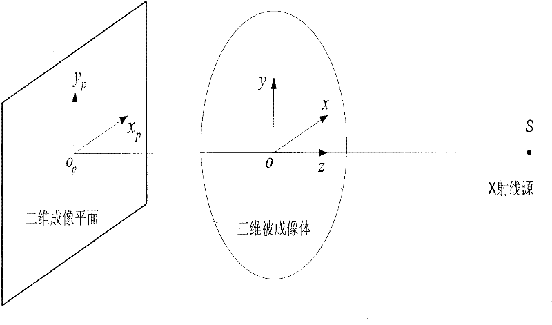
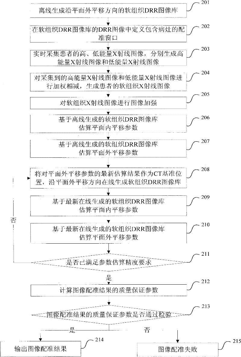
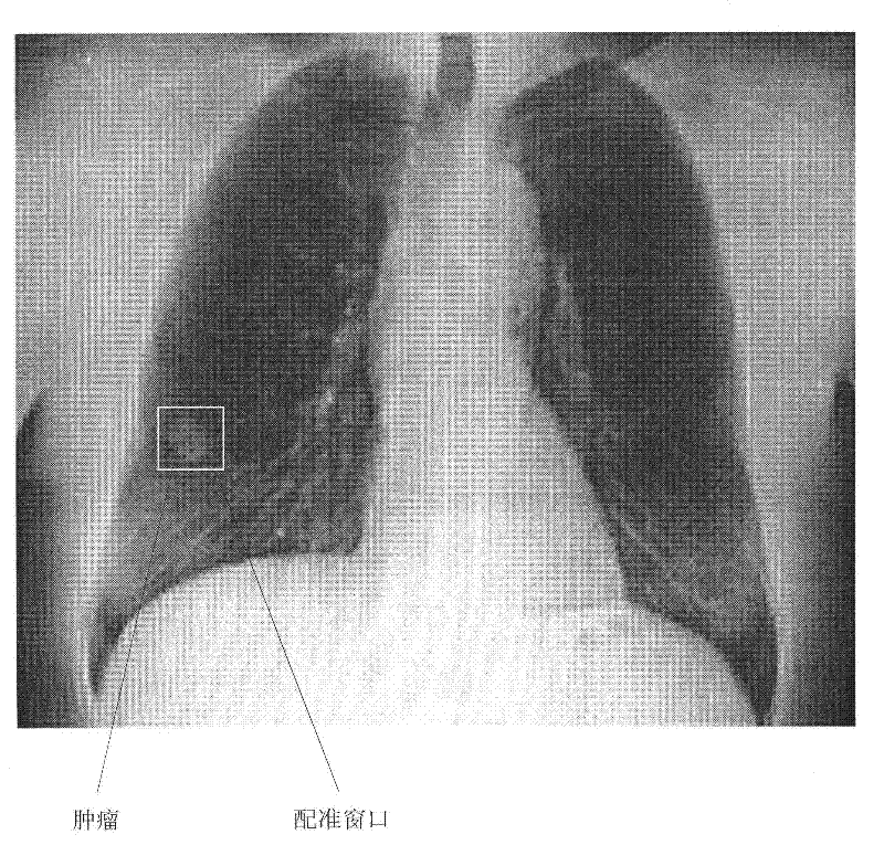
[0092] The core of the present invention is to generate a soft tissue DRR image library along the out-of-plane translation direction, and define a registration window containing a lesion (for example, a tumor) in the DRR image (which can be called a soft tissue DRR image) of the image library; The energy X-ray imaging technique generates a soft tissue X-ray image, and uses the soft tissue X-ray image as a registered image, and estimates the values ​​of the in-plane translation parameter and / or the out-of-plane translation parameter according to the registration window.

[0093] The above-mentioned dual-energy X-ray imaging technology refers to the continuous collection of two X-ray images of the same part of the human body with high and low energy X-rays. Due to the different attenuation coefficients of X-rays of different energies of human tissues, the two images will be different. The optical density distribution of the two images can be weighted and subtracted to give the di...


PUM


Abstract

The invention discloses a method and system for positioning a soft tissue lesion based on dual-energy X-ray images. The method comprises the following steps of: generating a three-dimensional image of a patient and offline generating a soft tissue DRR (Digitally Reconstructed Radiograph) image library along a plane inside translation direction; acquiring high- and low-energy X-ray images of the patient and generating a soft tissue X-ray image of the patient; taking the soft tissue X-ray image as a registered image and estimating values of a plane inside translation parameter and a plane outside translation parameter by using a registering window including the lesion in the DRR image in the soft tissue DRR image library generated offline; adjusting the three-dimensional image by a newest parameter estimation result for the plane outside translation parameter and generating the soft tissue DRR image library along the plane outside translation direction on line; and taking the soft tissue X-ray image as the registered image and further estimating the values of the plane inside translation parameter and the plane outside translation parameter by using the registering window including the lesion in the DRR image in the soft tissue DRR image library generated on line.

Description

technical field [0001] The invention relates to a medical image registration method and system, in particular to a method and system for locating soft tissue lesions based on dual-energy X-ray images. Background technique [0002] Image-guided radiation therapy (IGRT) is the latest theory and technology of tumor radiation therapy and tumor radiosurgery developed gradually in the past decade, and is a milestone of modern radiation therapy. IGRT uses advanced imaging equipment and image processing methods to locate and track patients' lesions before and during treatment, so as to achieve precise radiation therapy for tumors, reduce damage to normal tissues around tumors, and improve treatment effects for patients. IGRT is the basis for all modern emerging radiation therapy techniques such as stereotactic neuroradiosurgery (SRS), stereotactic body radiation therapy (SBRT), image-guided intensity-modulated radiation therapy (IG-IMRT), and image-guided IGRT core technology. [...

Claims


Application Information

Patent Timeline
IPC IPC(8): A61B6/00
Inventor 付东山
Owner 江苏瑞尔医疗科技有限公司
Learn More