Transdermal screw fixation in vitro sighting device for treating pelvic fracture

A pelvic fracture and aiming device technology, applied in the field of medical devices, can solve the problems of restricting the accuracy of navigation devices, reducing the accuracy of percutaneous nail placement, reducing the accuracy of nail placement, etc., and achieves shortened operation time, low price, and easy operation. Effect

Inactive Publication Date: 2012-07-18
THE AFFILIATED DRUM TOWER HOSPITAL MEDICAL SCHOOL OF NANJING UNIV
View PDF5 Cites 5 Cited by
  • Summary
  • Abstract
  • Description
  • Claims
  • Application Information

AI Technical Summary

Problems solved by technology

At present, the defects of this technology include: ① It is highly dependent on the 3D imaging navigation device, and the price of the device is usually more than 2 million yuan, which hinders its promotion in most hospitals; It takes time to synchronize the imaging data with the three-dimensional space information in the built-in computer of the navigation device, which is commonly referred to as "imaging registration". T...

Method used


Image

  • Transdermal screw fixation in vitro sighting device for treating pelvic fracture
  • Transdermal screw fixation in vitro sighting device for treating pelvic fracture
  • Transdermal screw fixation in vitro sighting device for treating pelvic fracture

Examples


Embodiment Construction

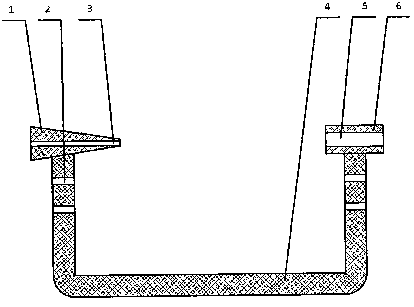
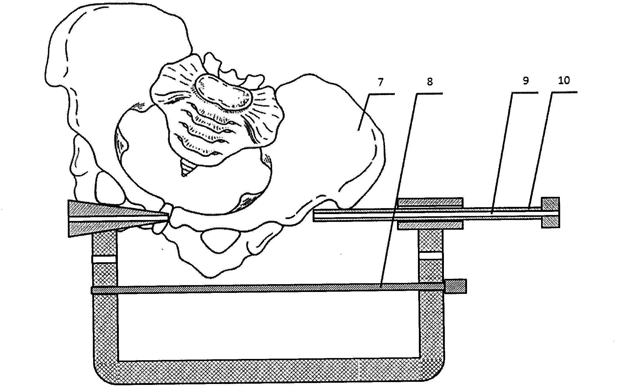
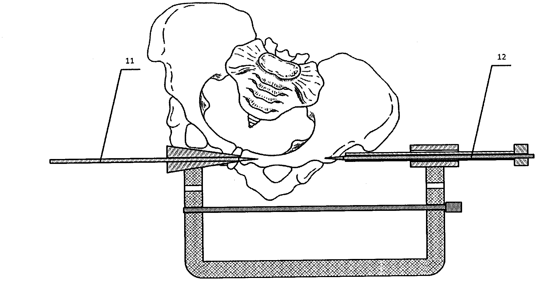
[0016] Such as figure 1 , figure 2 , image 3 , Figure 4 The structure of the invention shown:

[0017] The nail exit point sight 1 and the nail entry point sight 6 are respectively linked to the upper ends of the two ends of the U-shaped calibrated transparent frame 4, including the upper left end and the upper right end. The nail exit point sight 1 at the upper left end has a truncated cone shape and a wide left The narrow right part, the narrow right part is called the tip, the middle is the horizontal left guide needle channel 3, the nail point sight 6 at the upper right end is cylindrical in section, the middle is the horizontal inner sleeve channel 5, the left guide needle channel 3 and the inner The casing channel 5 is on the same horizontal plane; the upper left end of the U-shaped calibration transparent frame 4 has two calibration holes 2 from the nail exit sight 1 down, and they are all set on the left side of the U-shaped calibration transparent frame 4 Correspondin...


PUM


Abstract

The invention discloses a transdermal screw fixation in vitro sighting device for treating pelvic fracture. According to the sighting device, the left upper end of a U-shaped calibration transmissive frame is provided with a screw exit sight with a horizontal left guide needle passage in the middle, the right upper end of the U-shaped calibration transmissive frame is provided with a screw inlet sight with a horizontal inner sleeve passage in the middle, and calibration holes are respectively arranged on left and right sidewalls of the U-shaped calibration transmissive frame; a calibration rod is fixed on the U-shaped calibration transmissive frame through the calibration holes; an inner sleeve is inserted into the screw inlet sight through the inner sleeve passage; a left fixation guide needle is inserted into the left guide needle passage; and a right fixation guide needle is inserted into the inner sleeve through a right guide needle passage arranged in the inner sleeve. The invention solves the problems of high cost, long surgery time and low accuracy of a 3D imaging navigation device. The invention applicable to most hospitals has the advantages of simple operation process, shortened operation time, rapid adjustment of screw inlet and exit points and screw fixation axis, small volume, and convenient maintenance.

Description

Technical field [0001] The invention relates to a medical device, which is used in a set of operations, and particularly relates to a percutaneous nail placement external aiming device for pelvic fractures. Background technique [0002] The human pelvis has a relatively complex three-dimensional anatomical structure. Once a fracture occurs, surgical reduction and fixation are more difficult. Major surgical incisions are often required. It is difficult to meet the needs of treatment through extensive soft tissue exposure. [0003] Prior to the present invention, with the advancement of imaging technology, minimally invasive fixation of pelvic fractures with percutaneous nail placement has become a new development trend in surgical treatment of pelvic fractures. In the operation of this technology, expensive three-dimensional imaging navigation devices combined with fluoroscopy equipment are used to predict and monitor the position and direction of the nail to achieve accurate nail p...

Claims


Application Information

Patent Timeline
IPC IPC(8): A61B17/90
Inventor 施鸿飞熊进
Owner THE AFFILIATED DRUM TOWER HOSPITAL MEDICAL SCHOOL OF NANJING UNIV
Why Eureka
Try Eureka