Ultrasonic superficial tissue and organ volume scanning fracture imaging method

A scanning tomography and imaging method technology, applied in the field of medical imaging, can solve the problems of high cost, insufficient flexibility, and unsuitability for widespread promotion of a complete set of equipment, and achieve the effects of convenience, simplified requirements, and miniaturization

Inactive Publication Date: 2012-09-26
HUADONG HOSPITAL
View PDF5 Cites 11 Cited by
  • Summary
  • Abstract
  • Description
  • Claims
  • Application Information

AI Technical Summary

Problems solved by technology

Their common disadvantage is that the application flexibility is insufficient, and the cost of a complete set of equipment is high, which is unaffordable for many hospitals and is not suitable for widespread promotion.

Method used


Image

  • Ultrasonic superficial tissue and organ volume scanning fracture imaging method
  • Ultrasonic superficial tissue and organ volume scanning fracture imaging method
  • Ultrasonic superficial tissue and organ volume scanning fracture imaging method

Examples


Embodiment Construction

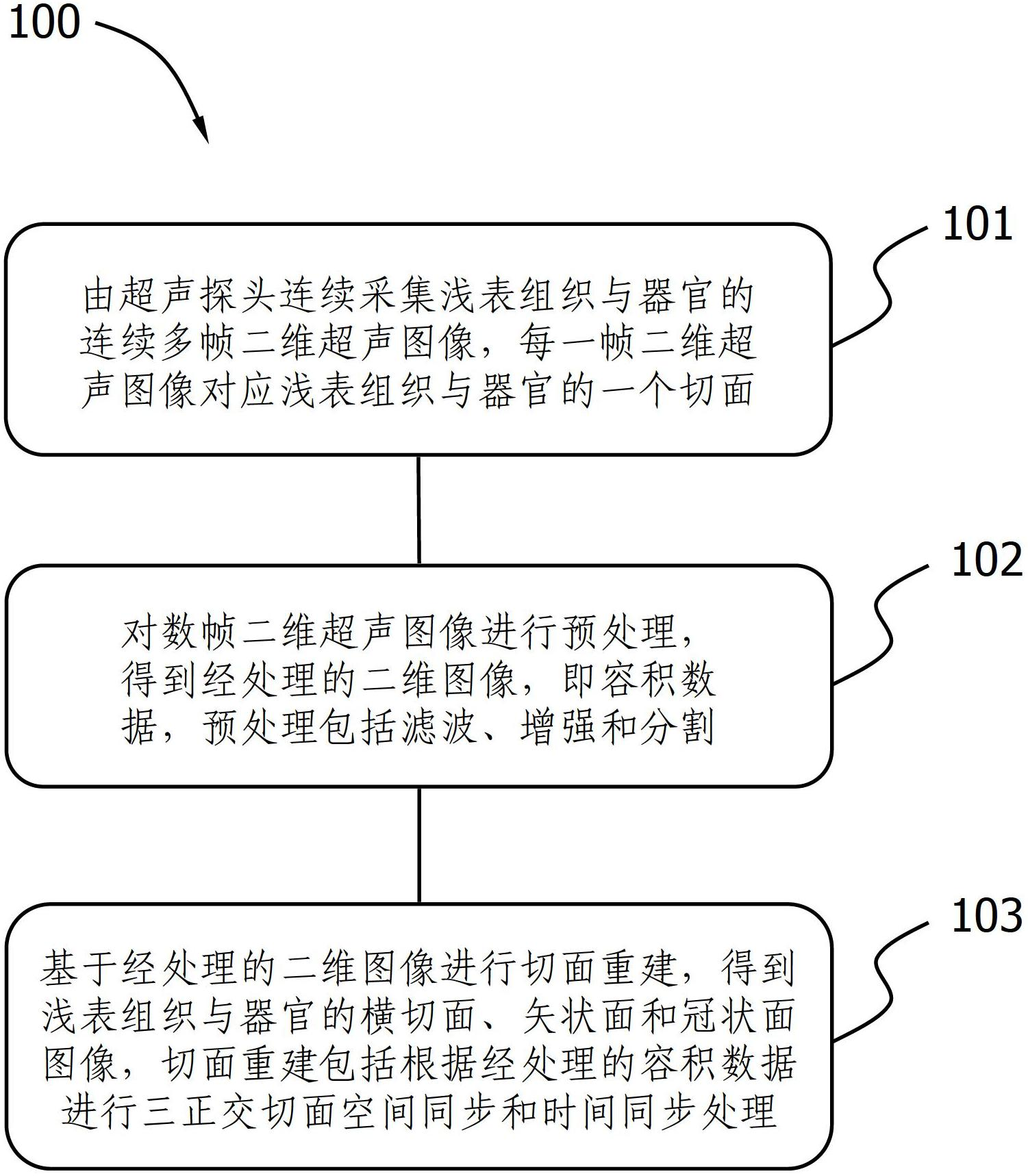
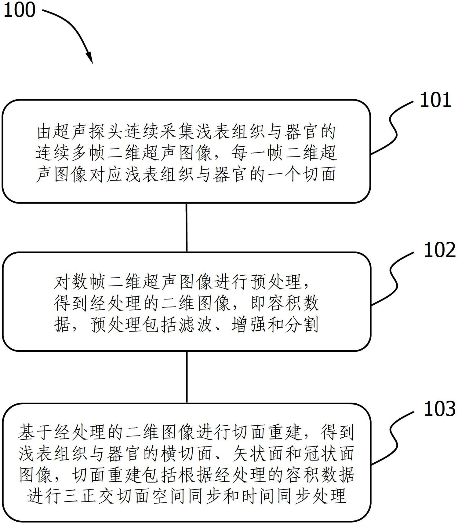
[0020] refer to figure 1 as shown, figure 1 Disclosed is a flow chart of an ultrasonic superficial tissue and organ volume scanning tomographic imaging method according to an embodiment of the present invention. The method 100 includes:

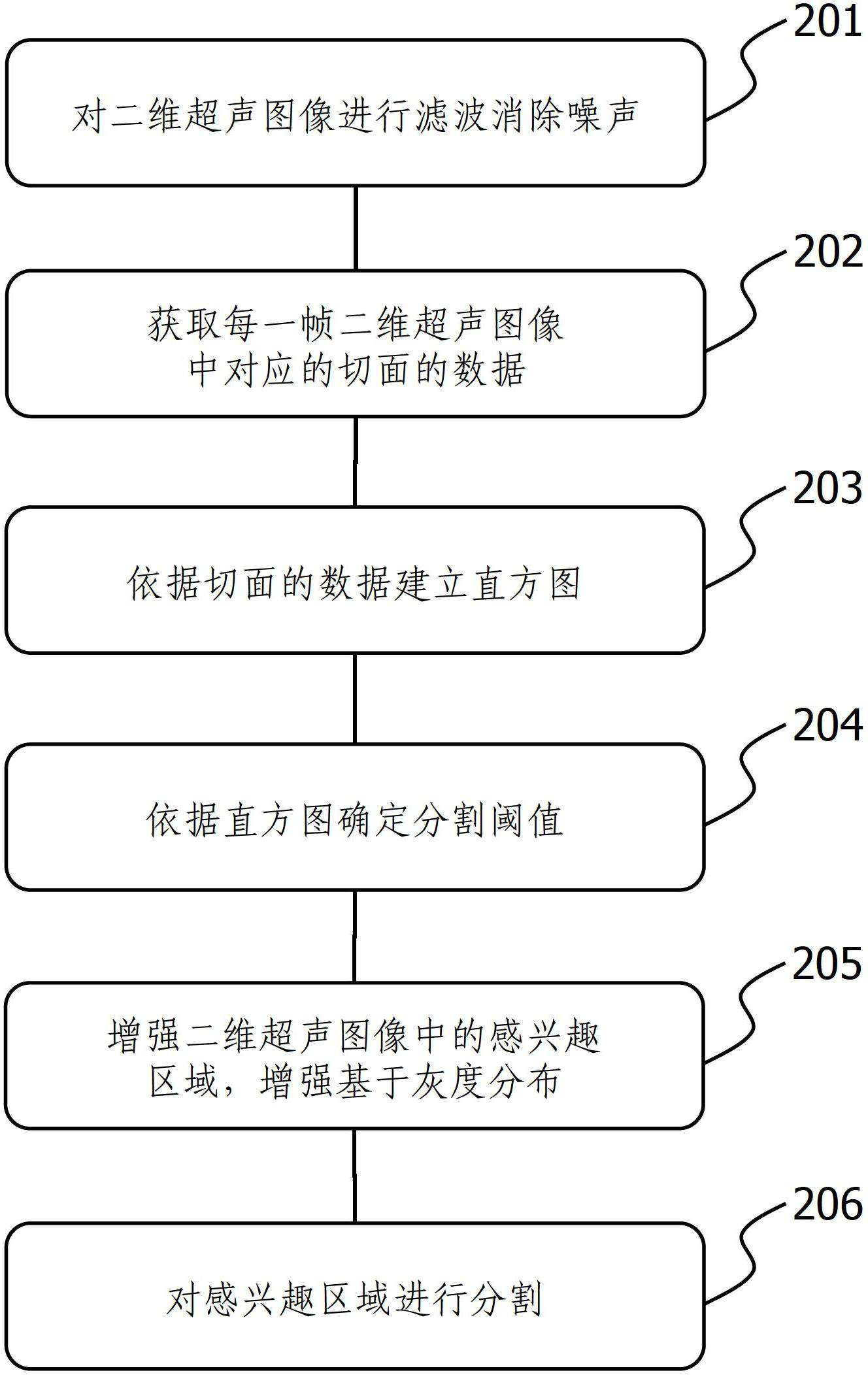
[0021] 101. Continuous multi-frame two-dimensional ultrasound images of superficial tissues and organs are continuously collected by the ultrasound probe, and each frame of two-dimensional ultrasound images corresponds to a section of superficial tissues and organs. In step 101, the present invention adopts two methods to obtain continuous two-dimensional ultrasound images. The first way is to use traditional mechanical means to control the ultrasound probe to move at a constant speed, and to collect images at a fixed interval frequency, such as 30 frames per second. Another way is to use equidistant sampling to collect images at fixed intervals through a scale and a series of calibration marks set at intervals on the scale, and make the de...


PUM


Abstract

The invention discloses an ultrasonic superficial tissue and organ volume scanning fracture imaging method, comprising the following steps of: collecting a continuous multi-frame two-dimensional ultrasonic image of the superficial tissue and organ by an ultrasonic probe, wherein each frame of two-dimensional ultrasonic image corresponds to a section of the superficial tissue and the organ; preprocessing the continuous multi-frame two-dimensional ultrasonic image to obtain the preprocessed volume data, wherein the preprocessing includes filtration, enhancement and division; reconstructing three sections of a cross section, a vertical plane and a coronal plane based on the processed two-dimensional image to obtain the multi-section image of the superficial tissue and the organ, wherein orthogonal section synchronous processing is carried out on the three-section image so that the cross section, the vertical plane and the coronal plane perform are subjected to time synchronization and space synchronization, and the three orthogonal section synchronous processing includes time synchronous processing and space synchronous processing. By utilizing a regular superficial linear array probe, the fracture imaging process is simple, and the superficial tissue and organ volume scanning fracture imaging device is miniaturized and convenient.

Description

technical field [0001] The invention relates to medical imaging technology, in particular to an ultrasonic superficial tissue and organ volume scanning tomographic imaging method. Background technique [0002] Ultrasound (Ultrasound) medicine is a discipline that combines acoustics, medicine, optics and electronics. Ultrasound medicine is the study of the application of acoustic technology higher than audible sound frequency in the medical field. Including ultrasonic diagnostics, ultrasonic therapeutics and biomedical ultrasonic engineering, ultrasonic medicine has the characteristics of combining medicine, science and engineering, involves a wide range of contents, and has high value in the prevention, diagnosis and treatment of diseases. [0003] An important item in ultrasound medicine is ultrasound imaging technology. Ultrasound imaging technology is to transmit ultrasonic waves by a probe and receive the reflected ultrasonic waves. An image is generated based on the ...

Claims


Application Information

Patent Timeline
Patent Type & Authority Applications(China)
IPC IPC(8): A61B8/13
Inventor 陈悦陈林
Owner HUADONG HOSPITAL
Why Eureka
Try Eureka