Magnetic resonance imaging-based perforator flap blood vessel positioning and measurement method

A magnetic resonance imaging and blood vessel technology, which is used in diagnostic recording/measurement, medical science, diagnosis, etc., can solve the problems of inaccurate positioning and cannot reflect the length and diameter of perforating vessels, and achieve accurate positioning, less time, and efficiency. high effect

Active Publication Date: 2012-11-07
EAST CHINA NORMAL UNIV
View PDF7 Cites 10 Cited by
  • Summary
  • Abstract
  • Description
  • Claims
  • Application Information

AI Technical Summary

Problems solved by technology

The disadvantage of this method is that the positioning is not accurate, and it cannot reflect the length and diameter of the perforating vessels.

Method used


Image

  • Magnetic resonance imaging-based perforator flap blood vessel positioning and measurement method
  • Magnetic resonance imaging-based perforator flap blood vessel positioning and measurement method
  • Magnetic resonance imaging-based perforator flap blood vessel positioning and measurement method

Examples


Embodiment

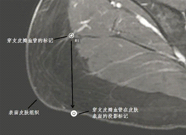
[0038] The magnetic resonance image data collected in this embodiment is a contrast agent-enhanced T1-weighted image of the abdomen of a human body, and the data comes from a GE1.5T magnetic resonance imager.

[0039] Step 1: Acquisition of a contrast agent-enhanced T1-weighted magnetic resonance image of human subcutaneous tissue by using magnetic resonance imaging technology. The imaging sequence used is a gradient echo sequence with fat signal suppressed, and its imaging parameters are as follows: recovery time is 6.064 ms, echo time is 2.92 ms, dump angle is 15 degrees, observation field is 480*480 mm, slice thickness is 3mm, the scanning matrix is ​​512*512. The image display tool used is OsiriX software.

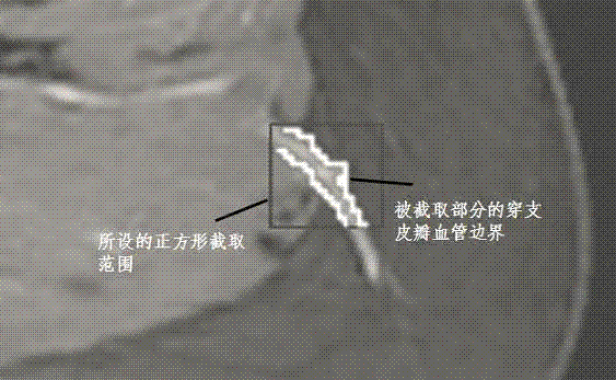
[0040] Step 2: Select a reference point and mark the perforator flap vessels.

[0041] In this embodiment, the navel in the abdomen of the human body is selected as the reference point, and three perforating flap vessels are found and marked on the left side of the a...


PUM


Abstract

The invention discloses a magnetic resonance imaging-based perforator flap blood vessel positioning and measurement method, which comprises the following steps of: acquiring a magnetic resonance image of human subcutaneous tissues, and selecting a reference point from the image; searching for a position where a perforator flap blood vessel penetrates a muscle in the image, and setting mark points; reading imaging coordinates of the reference point and the mark points, and calculating displacement between the reference point and each mark point; tracking and segmenting the blood vessel, and calculating the length and diameter of the perforator flap blood vessel; and projecting all the mark points onto the surface of skin to obtain a perforator flap blood vessel mark image on the surface of the skin. The method can be used for accurately positioning the perforator flap blood vessel and accurately measuring the length and diameter of the perforator flap blood vessel, and is short in manual operation time and high in efficiency.

Description

technical field [0001] The method of the invention belongs to the technical field of magnetic resonance imaging, and in particular relates to a method for positioning and measuring perforator flap blood vessels based on magnetic resonance imaging. Background technique [0002] Perforating vessels refer to small, stem-like vessels that connect and penetrate into muscle tissue, supplying blood to flap tissue and regenerating subcutaneous tissue. Perforator flap refers to a large flap tissue composed of skin and fat tissue supplied by perforator vessels. In today's clinical plastic surgery, perforator flap surgery has become more and more widely used, such as skin grafting, breast reconstruction surgery and so on. Accurate measurement of the position and diameter of perforator flap vessels can provide surgeons with effective anatomical information, which is beneficial to the perforator flap surgery. [0003] The traditional perforator flap blood vessel location method is to u...

Claims


Application Information

Patent Timeline
Patent Type & Authority Applications(China)
IPC IPC(8): A61B5/055A61B5/107
Inventor 王丽嘉裴孟超李建奇王乙
Owner EAST CHINA NORMAL UNIV
Why Eureka
Try Eureka