Magnetic resonance imaging method

A magnetic resonance imaging and imaging technology, applied in the fields of medical science, sensors, diagnostic recording/measurement, etc., can solve the problem that cannot fully meet the requirements of tissue and organ discrimination and localization, poor adipose tissue signal suppression effect, and poor tissue imaging quality, etc. problem, to reduce image artifacts, accurate and reliable results, and improve imaging quality.

Active Publication Date: 2012-11-14
SHENZHEN UNITED IMAGING HEALTHCARE CO LTD
View PDF3 Cites 24 Cited by
  • Summary
  • Abstract
  • Description
  • Claims
  • Application Information

AI Technical Summary

Problems solved by technology

[0006] The purpose of the present invention is to provide a magnetic resonance imaging method, which is used to solve the problem that the existing adipose tissue signal suppression effe...

Method used


Image

  • Magnetic resonance imaging method
  • Magnetic resonance imaging method
  • Magnetic resonance imaging method

Examples


Embodiment Construction

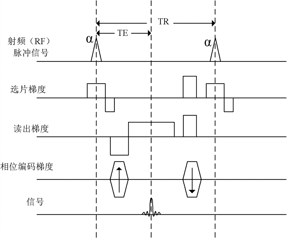
[0024] As mentioned above in the background technology: figure 1 It is a schematic diagram of a common FLASH imaging pulse sequence, by applying radio frequency RF pulse signals and gradient pulses (including film selection gradients, phase encoding gradients and readout gradients) to the object under inspection in a static magnetic field, and giving echo signal acquisition The tissue image corresponding to the object to be inspected is reconstructed from the magnetic resonance (Magnetic Resonance, MR) signal emitted by the protons in the inspected area by means of a method. In ordinary FLASH sequence applications, the signal of fat is relatively high, which often covers the diseased tissue, which is not conducive to diagnosis; in addition, because the precession frequency of hydrogen protons in human body fat and water is different (the precession frequency of water protons is slightly faster For fatty protons), it is easy to cause chemical shift artifacts and affect the qual...


PUM


Abstract

The invention provides a magnetic resonance imaging method for dynamic contrast-enhanced imaging based on a three-dimensional pulse sequence of fast low angle shot FLASH. The method comprises the steps of: applying a FLASH imaging pulse sequence to an objected to be examined in a static magnetic field, wherein the FLASH imaging pulse sequence comprises radio frequency shot pulses a and fat inhibiting pulses b, and N radio frequency shot pulses a are comprised between two adjacent fat inhibiting pulses b; adjusting a fat inhibiting flip angle b with certain time of inversion T1 and time of scan delay TD to obtain the optimal fat inhibiting effect; and adjusting the time of inversion T1 and/or the time of scan delay TD to obtain the optimal fat inhibiting effect in a condition that the optimal fat inhibiting effect cannot be obtained by adjusting the fat inhibiting flip angle b. Compared with the prior art, the magnetic resonance imaging method provided by the invention has the advantages of good fat inhibiting effect and high imaging quality.

Description

technical field [0001] The invention relates to a magnetic resonance imaging technique, in particular to a magnetic resonance imaging method for suppressing fat tissue signals by applying fat suppression pulses. Background technique [0002] Magnetic resonance imaging (Magnetic Resonance Imaging, MRI) is such a technique: by applying a gradient magnetic field and RF waves to the object under inspection in a static magnetic field, and giving magnetic resonance ( Magnetic Resonance (MR) signals to reconstruct the structural image corresponding to the object being inspected. [0003] Applying this technique to imaging the internal structures of the human body has resulted in a revolutionary medical diagnostic tool. The application of rapidly changing gradient magnetic fields has greatly accelerated the speed of nuclear magnetic resonance imaging, making the application of this technology in clinical diagnosis and scientific research a reality, and greatly promoting the rapid d...

Claims


Application Information

Patent Timeline
IPC IPC(8): A61B5/055
Inventor 张强
Owner SHENZHEN UNITED IMAGING HEALTHCARE CO LTD
Why Eureka
Try Eureka